Armamentarium
|
History of the Procedure
The French anatomist René Le Fort published his classic treatise on the description of common fracture patterns in the middle facial third in 1901. The ensuing engulfment of the World Wars produced horrendous mass casualties that steered facial reconstructive surgeons like Kazanjian in their efforts to manage injuries to the middle third of the face. Building on the knowledge and skill born by these conflicts, Sir Harold Gillies, an otolaryngologist by training, was the first surgeon to publish an attempt at mobilization of the midface in the management of a patient with craniofacial dysostosis. The procedure was unsuccessful, and Gillies later abandoned it. Subsequently, Longacre attempted reconstruction of the midface by autogenous rib grafting. However, this procedure did nothing to address the functional impairments associated with total midface deficiency. In addition, the long-term stability of the reconstruction from an aesthetic standpoint was questionable. In 1967, the pioneering efforts of Tessier revolutionized management of the patient with total midface deficiency. His landmark presentations and publications concerning the mobilization of the entire middle face through the concept of combined intra- and extracranial approach in a safe and consistent manner were groundbreaking. Modifications and extensions of this concept by Tessier himself and others have produced surgical techniques that have provided relief of functional impairments and improved the stability of facial aesthetics to the benefit of the patient with total midface deficiency. The first known performance of this surgery in the United States was by Robert V. Walker in 1967, shortly after Tessier’s presentation in Rome.
Indications for the Use of the Procedure
Craniofacial anomalies, by their very nature, are repetitive patterns of deformity affecting the different functional and aesthetic subunits of the facial hard and soft tissues. Perhaps their only common feature is the degree of variable expressivity of the deformity within each subtype of anomaly. The surgical management of the full spectrum of craniofacial anomalies affecting the subcranial facial skeleton is beyond the scope of this chapter.
Craniofacial Dysostosis
The craniofacial dysostosis syndromes—Apert, Crouzon, Pfeiffer, and Saethre-Chotzen—are characterized by sutural involvement that not only includes the cranial vault but also extends into the skull base and midfacial skeletal structures. Although the cranial vault and cranial base are thought to be the regions of primary involvement, there is also significant impact on midfacial growth and development. In addition to cranial vault dysmorphology, patients with these inherited conditions exhibit a characteristic “total midface” deficiency that must be addressed as part of the staged reconstructive approach. Although there is some similarity between the pattern of facial growth and development in these patients, there is a high degree of variable expressivity in each patient regardless of syndrome. This must be taken into account when planning and executing surgical correction of these deformities.
Total Midface Deficiency
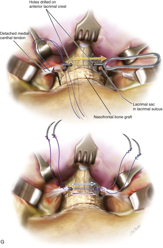
The role of the human face is significant in both a direct and an indirect fashion for reasons other than purely aesthetic considerations. These are secondary to the highly evolved and specialized functions of the face in vision, breathing, speech production, smell, and hearing, among a few. In patients with craniofacial dysostosis, in addition to potential neurologic deficits, there is often variable fusion of the lesser sutures of the skull base. This commonly results in abnormal ophthalmologic findings including exorbitism, exotropia, orbital dystopia, and ptosis secondary to a lack of orbital depth and diameter, as well as prolapse of the ethmoid sinuses through the medial orbital walls. The severe occlusal discrepancies found in this group of patients are characterized by generalized hypoplasia of the maxilla, transverse deficiency, class III malocclusion, and apertognathia. All of these abnormalities contribute to impair speech articulation and mastication. In addition, cleft palate, when present, can produce velopharyngeal incompetence. The severe retrusive position of the midface can also interfere with nasal breathing and produce chronic nasal obstruction. Varying degrees of orbital hypertelorism (OHT) may or may not be present. The extent to which this is present strongly influences the type of surgical correction required for total midface deficiency ( Figure 46-1, A-H ).
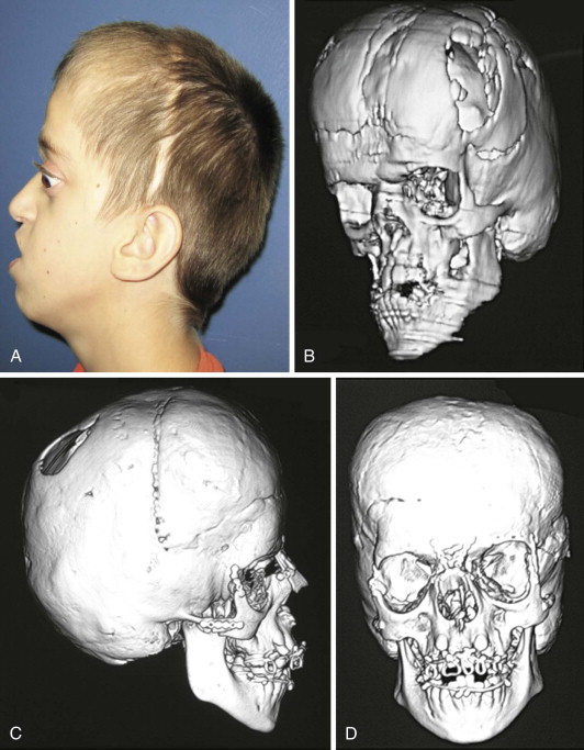
The presence of total midface deficiency does not mitigate the coexistence of other facial skeletal abnormalities such as mandibular excess and retrogenia. In addition, there are often irregularities of the forehead as well as frontal bossing. Typically the nasal length is short and projection is deficient. This gives rise to an exaggerated depression or flatness of the nasofrontal region. Because of the deficiency in orbital depth and diameter producing an exorbita, an excessive amount of scleral show may be present. Ptosis of the lids and lateral canthal dystopia are usually present. The nasolabial angle is commonly less than 90 degrees because of nasal deficiency and the overprojection of the maxillary teeth.
A more precise measurement of the degree of exorbitism can be obtained using the Hertel exophthalmometer or through computed tomography (CT) scan analysis of the position of the globe relative to the eye and the upper facial skeletal to normative values as originally published by Posnick and colleagues.
Limitations and Contraindications
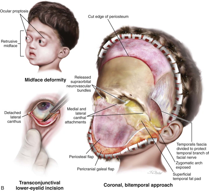
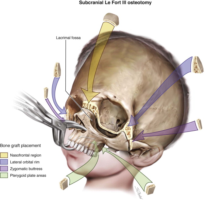
However, specific limitations apply to the use of this surgical maneuver. The bony structures of the middle facial third are in contiguity with the cranial base superiorly. Transgression of this natural barrier is called for in the surgical correction of some craniofacial anomalies. If the presenting deformity extends to excessive interorbital distance or there is significant aberration of the supraorbital/forehead subunit, consideration should be given to using a combined intra- and extracranial approach such as facial bipartition or monobloc osteotomy. In addition, subcranial Le Fort III osteotomy does not address the three-dimensional vertical slanting of the facial halves or the convex arc of rotation of the face, as seen in some craniofacial dysostosis patients, which can only be adequately managed with facial bipartition osteotomy.
The surgeon must give thoughtful consideration to addressing the presenting dysmorphology, with the intention of improving the overall aesthetic and functional concerns of the patient while taking into account the potential complications and benefits inherent with the use of an intracranial approach. Management of the skeletally immature patient, as discussed later, requires surgical intervention based on the complex balance between the long-term stability of the correction and more immediate functional, psychological, and aesthetic demands of each patient. As with all craniofacial procedures, patient selection is critical for a successful outcome.
Technique: Le Fort III Osteotomy
Step 1:
Intubation
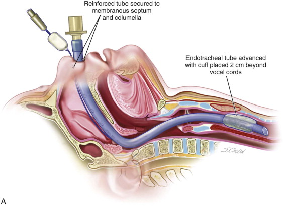
Nasoendotracheal intubation with a reinforced tube exiting inferiorly across the mouth, neck, and chest before returning to anesthesia is preferred. The tube is secured with suture to the membranous septum and columella. Because intermaxillary fixation is necessary to establish the projection of the middle face, oral intubation is less desirable and should be avoided unless the splint can be modified to accommodate the position of the tube. The length of the endotracheal tube must be sufficiently below the level of the vocal cords to prevent unintended dislodgment during midface disimpaction and advancement ( Figure 46-2, A ).
Stay updated, free dental videos. Join our Telegram channel
VIDEdental - Online dental courses


