Chapter 14
Ectopic eruption of maxillary permanent canines
Jane A. Soxman
The maxillary permanent canine normally erupts at the age of 11–12 years (American Academy of Pediatric Dentistry, 2014). The canine bulge should be palpable high in the alveolar process above the primary canine by the age of 9–10 years if dental and chronological ages are synonymous. At the age of 11–12 years, the absence of the canine bulge indicates a significantly higher possibility of palatal displacement (Chalakkal et al., 2011). Digital palpation to determine the status of the maxillary permanent canines should be routinely performed. If the canine bulge is not palpated or the canine appears to be in an abnormal position with mesial inclination on palpation, radiographic examination with a panoramic or periapical radiograph is indicated. Abnormal inclination of the canine, mesially angled toward or superimposed over the root of the permanent lateral incisor or premolar, signals the need for immediate intervention to avoid root resorption, which can rapidly occur (Fricker et al., 2008) (Figures 14.1–14.3a, 14.3b, and 14.4).
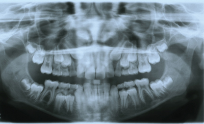
Figure 14.1 Panoramic radiograph showing bilateral ectopic position of maxillary permanent canines.
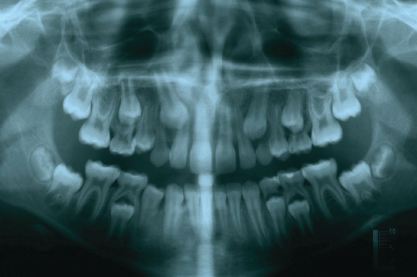
Figure 14.2 Panoramic radiograph showing abnormal inclination of the maxillary right permanent canine superimposed over the root of the maxillary right permanent lateral incisor.
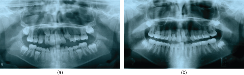
Figure 14.3 (a) Panoramic radiograph showing ectopic position maxillary right permanent canine. (b) Panoramic radiograph showing root resorption of the maxillary right permanent lateral incisor resulting from ectopic eruption of the maxillary right permanent canine.
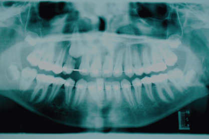
Figure 14.4 Panoramic radiograph showing root resorption of the maxillary right first premolar due to ectopic eruption of the maxillary right permanent canine.
An overretained maxillary primary canine may indicate ectopic position of a permanent maxillary canine or a congenitally missing permanent successor (Figures 14.5a, 14.5b, 14.6a, and 14.6b).
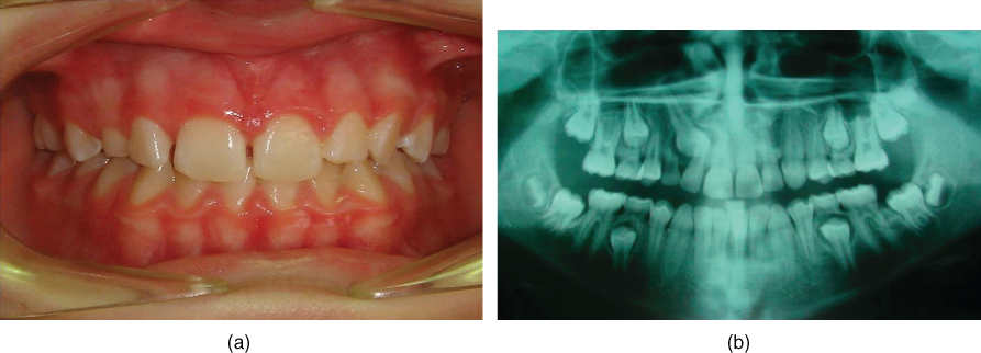
Figure 14.5 (a) Photograph showing overretained maxillary right primary canine. (b) Panoramic radiograph showing ectopic position of maxillary right permanent canine with overretained maxillary right primary canine.
Stay updated, free dental videos. Join our Telegram channel
VIDEdental - Online dental courses


