Armamentarium
|
History of the Procedure
The earliest attempts at cleft management used simple coaptation of the margins of the cleft after they had been surgically exposed. The results obtained with these techniques were unfortunately consistently bad, for many reasons, but particularly because of poor surgical reconstruction of the labial musculature. It was on muscle reconstruction that Victor Veau concentrated in his book, Cleft Lip, Clinical-Surgical Forms, published in 1938. Through the principles espoused by Veau and more recently by Delaire , modern cleft management has come to recognize the importance of (and to incorporate into surgical approaches) (1) wide undermining by surgical dissection of the muscles of the lip, sill, and floor of the nose; (2) detailed reconstruction of the mucoperiosteal plane on the nasal side of the primary palate; (3) solid anchorage of the muscles of the nasal floor and the sill to the region of the anterior nasal spine; and (4) progressive and systematic suturing of the orbicularis muscles of the upper lip, which have been surgically defined on both sides of the cleft.
Indications for the Use of the Procedure
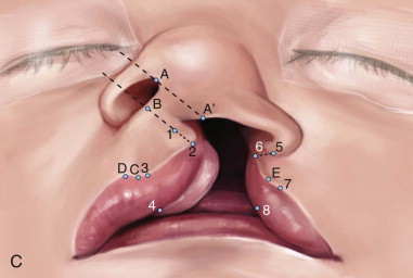
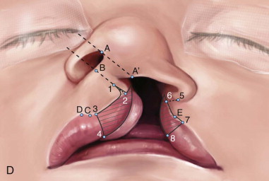
Congenital labiomaxillary clefts result from the absence of fusion or from incomplete fusion of the maxillary and medial nasal processes. The superficial muscles of the face, which arise from the second branchial arch, migrate laterally and medially between the epidermis and subjacent ectomesenchyme and normally reach the midline in the week after fusion of the facial processes. In a complete labiomaxillary cleft, the muscles of the nasal floor and the upper lip cannot bridge the gap of the cleft, nor can they unite with their muscular counterparts on the noncleft side. The muscular integrity of the region is considerably disrupted, which has a profound effect on the underlying skeleton. Abnormal muscle architecture is present during bone formation, and virtually all bone formation in the cleft fetus takes place at the direction and under the influence of dyssymmetric and distorted muscle forces. To achieve the proper midline position and attitude of the nasal septum, the surgeon must perform a wide subperichondrial dissection of the cleft side of the septum. This permits the creation of a watertight nasal floor while preserving the integrity of the maxillary labial frenum, which, as described previously, is an important constituent of the septopremaxillary traction system. It is necessary to reconstruct the nasolabial muscles of the cleft so that the result is a symmetric influence on the nasal septum from both the cleft and noncleft muscles. Successful lip/nose repair must establish a straight nasal septum positioned in the facial midline; symmetric reconstruction of the nasolabial muscles; absence of a vestibular oral-nasal communication; and a functional patent nostril on the cleft side, all of which are essential for good subsequent facial growth.
Technique: Functional Cheilorhinoplasty of Delaire
Step 1:
Surgical Approach
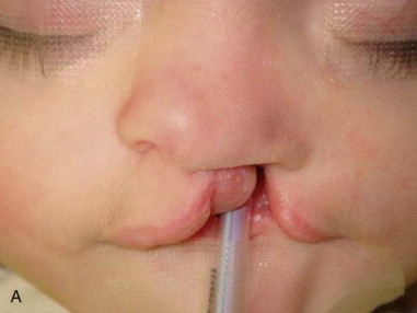
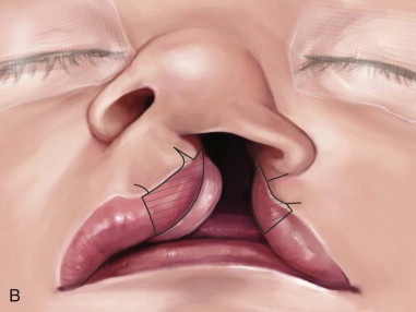
At the beginning of the operation, the surgeon must adopt a surgical approach that is based on functional anatomy. The surgeon must avoid following a geometric plan ( Figure 50-1, A ).
Stay updated, free dental videos. Join our Telegram channel
VIDEdental - Online dental courses


