Temporomandibular joint ankylosis is a debilitating condition characterized by abnormal bony fusion within the joint, resulting in restricted jaw movement, facial asymmetry, and functional impairment. It commonly arises from trauma but may also result from infection, inflammation, or systemic factors. Diagnosis relies on detailed imaging, via computed tomography scans, to guide surgical planning. Management requires careful airway considerations and a structured surgical approach, with osteoarthrectomy forming the cornerstone of treatment. Reconstruction aims to restore mandibular height and function using techniques such as costochondral grafting, distraction osteogenesis, or total joint replacement, selected based on patient age, severity, and recurrence risk.
Key points
-
•
Temporomandibular joint ankylosis requires customized management by an experienced surgeon and anesthetist.
-
•
Airway security : Flexible bronchoscopic-guided nasotracheal intubation is preferred. Total intravenous anesthesia without neuromuscular blockade is used to enable facial nerve monitoring and minimize the risk.
-
•
Surgical approaches : Preauricular-endaural or extended temporal incision minimizes scarring and temporal facial nerve injury, and combined with retromandibular approach for total joint replacement.
-
•
Osteoarthrectomy with total joint replacement is the adult gold standard and is increasingly used after failed pediatric arthroplasty, although autogenous reconstruction remains preferred in children.
-
•
Postoperative care : Aggressive mouth opening physiotherapy is essential for sustained mouth opening and preventing reankylosis.
Abbreviations
| COS | concomitant orthognathic surgery |
| CT | computed tomography |
| MIO | maximum interincisal opening |
| OPG | orthopantomogram |
| PJI | prosthetic joint infection |
| PSI | patient-specific implant |
| RCU | ramus condyle unit |
| SCM | sternocleidomastoid muscle |
| STL | stereolithographic |
| TJR | total joint replacement |
| TMJA | temporomandibular joint ankylosis |
| VSP | virtual surgical planning |
Introduction
Temporomandibular joint ankylosis (TMJA) involves heterotopic ossification in the joint space between the mandibular condyle and glenoid fossa, leading to restricted mandibular mobility. This end-stage TMJ disease impairs mandibular growth and function, reducing quality of life. The most common etiology is trauma (79%–90%), followed by infection spreading from middle ear, inflammatory arthritis of TMJ, late degenerative joint disease (DJD) and unexplained causes, among others. ,
In unilateral TMJA , the shortened ramus and mandibular body manifest as facial asymmetry, chin deviation, occlusal cant, and reduced posterior airway space. The severity depends on age of onset and duration of ankylosis. Long standing bilateral involvement would cause severe retrognathia. Restricted contralateral laterotrusion results from bony fusion and atrophy of the ipsilateral lateral pterygoid muscle ( Fig. 1 A-D ).
( A ) Preoperative frontal photograph of a 10 years old male with deviated chin and facial asymmetry due to left TMJA. ( B ) Preoperative frontal photograph of a pediatric female patient with mandibular retrognathia due to bilateral TMJA. ( C ) Preoperative profile photograph of the patient in Fig. 1 B showing mandibular retrognathia. ( D ) Profile photograph of the same patient showing mouth opening and progressive mandibular retrognathia at 10 years follow-up after gap arthroplasty.
Imaging modalities of choice are computed tomography (CT; cone-beam or noncontrast, 0.6–1 mm slice thickness, zero-degree gantry tilt) and orthopantomogram (OPG). Consider CT angiogram in recurrent cases with large ankylotic mass to assess proximity/entrapment of the maxillary artery (risk of hemorrhage; may require preoperative embolization or intraoperative external carotid ligation).
-
•
Evaluate glenoid fossa integrity and temporal bone pneumatization.
-
•
Detailed structured reporting aids surgical planning.
Additional radiographic features include enlarged ipsilateral coronoid process and deepened antegonial notch due to unopposed activity of ipsilateral temporalis, masseter, and medial pterygoid muscles.
Anesthetic Considerations : Reduced mouth opening and associated severe facial deformity poses an anesthetic challenge. Preferred technique is flexible fibreoptic bronchoscopy guided nasotracheal intubation in spontaneously breathing patients (awake or sedated). Topical airway anesthesia via nebulization, nerve blocks, or spray-as-you-go. Extend safe apnoea time with apnoeic oxygenation (nasal cannula or nasopharyngeal airway); advanced options include high-flow nasal oxygenation or THRIVE.
Management philosophy
Ankylosis should be released as soon as it is diagnosed. The mainstay of management is radical release of ankylosis (1–1.5 cm gap), and ramus condyle unit (RCU) reconstruction with costochondral graft (CCG) in pediatric patients, transport disk distraction osteogenesis (TDDO) in pediatric/adult patients, and total joint replacement (TJR) in adults followed by aggressive physical therapy (PT). Treatment is tailored to the patients age, duration and severity of ankylosis, associated deformity and requirement of future growth. Strategies for mitigation of reankylosis include interposing a barrier between temporal bone and cut end at ramus by temporalis muscle/fascia flap, fat packing and aggressive PT. Many of long standing TMJA patients will also have associated obstructive sleep apnea (OSA) due to retrognathia. A mandibular distraction should be performed before release. This increases the posterior pharyngeal airway space. In adults TJR with concomitant orthognathic surgery employing counterclockwise rotation of maxillomandibular complex is helpful.
Criteria for selection of CCG, TDDO, or total joint replacement in temporomandibular joint ankylosis for adults and growing patients
-
•
The choice between CCG, TDDO, or TJR in the management of TMJ ankylosis depends primarily on age/growth status, extent of mandibular deformity, previous surgeries, condition of the joint and surrounding tissues, and expected functional outcomes ( Table 1 , Fig. 2 ).
Table 1
Table showing decision-making framework based on various clinical factors
Clinical factor Costochondral Graft Transport Disk Distraction Osteogenesis Total Joint Replacement Growing patient (<12 y) Preferred Useful in selected cases Generally contraindicated except in multiple operated cases Adult patient Less predictable Selected cases Preferred option Need for growth potential Yes Yes but limited No Recurrent ankylosis (pediatric or adult) Not favorable Favorable Strong indication Multiple previous surgeries (pediatric or adult) Not favorable Favorable Strong indication Severe RCU deficiency Good in pediatric Good in pediatric/adult Excellent in pediatric and adults Bilateral ankylosis Good in pediatric Technically demanding Excellent in adults Need for immediate function Moderate in pediatric Good in pediatric & adult Excellent Predictability Variable in pediatric/adult Variable in pediatric/adult Highest Donor-site morbidity Present None None Fig. 2 Flowchart showing treatment modality for RCU reconstruction in temporomandibular joint ankylosis patients.
Contemporary consensus
-
•
Children: CCG remains the standard reconstructive option for first time operation because of its growth potential.
-
•
Young patients with severe ramus deficiency: TDDO is an attractive biological alternative only in experienced hands.
-
•
Adults, especially recurrent ankylosis and failed reconstructions: TJR provides the most predictable functional and skeletal outcome and is increasingly regarded as the gold standard for TMJ ankylosis reconstruction.
-
•
Multiple failed arthroplasty in children or adults require TJR for restoration of function.
Single stage vs 2-stage reconstruction
Selecting between a single-stage (resection and immediate joint reconstruction) and 2-stage (resection first, followed by delayed reconstruction) approach depends largely on patient age, the severity of the ankylosis, and anatomic complexities.
Single stage is preferred for adult cases (a) without extensive bony ankylosis, (b) have acceptable facial aesthetics before surgery, (c) who can maintain stable occlusion on CT imaging and, (d) fossa anatomy that requires only minor modifications ( Box 1 ).
Box 1
Box showing Single-stage protocol for reconstruction in temporomandibular joint ankylosis
-
Single-stage protocol (adults with favorable anatomy)
-
Sequence:
-
Preoperative Planning → Single Operation → Immediate Recovery
-
-
Stage Details:
-
•
Step 1: Preoperative Planning: Take high-resolution CT scans. Perform 3D virtual surgery to plan bone cuts. Manufacture custom cutting templates and then finalize TJR.
-
•
Step 2: Operation: Perform a single surgery-osteoarthrectomy using the custom guides. Immediately fix the final custom joint and secure the planned occlusion.
-
•
Step 3: Postoperative Care: Start aggressive PT within 24 hours.
-
•
-
Targets:
-
•
Patient Age: ≥14 to 16 year old
-
•
On table Maximum Interincisal Opening (MIO) Target: greater than 35 mm passive opening on the operating table
-
•
Postoperative MIO Target: 35 mm to 40 mm maintained at 6 to 12 months
-
•
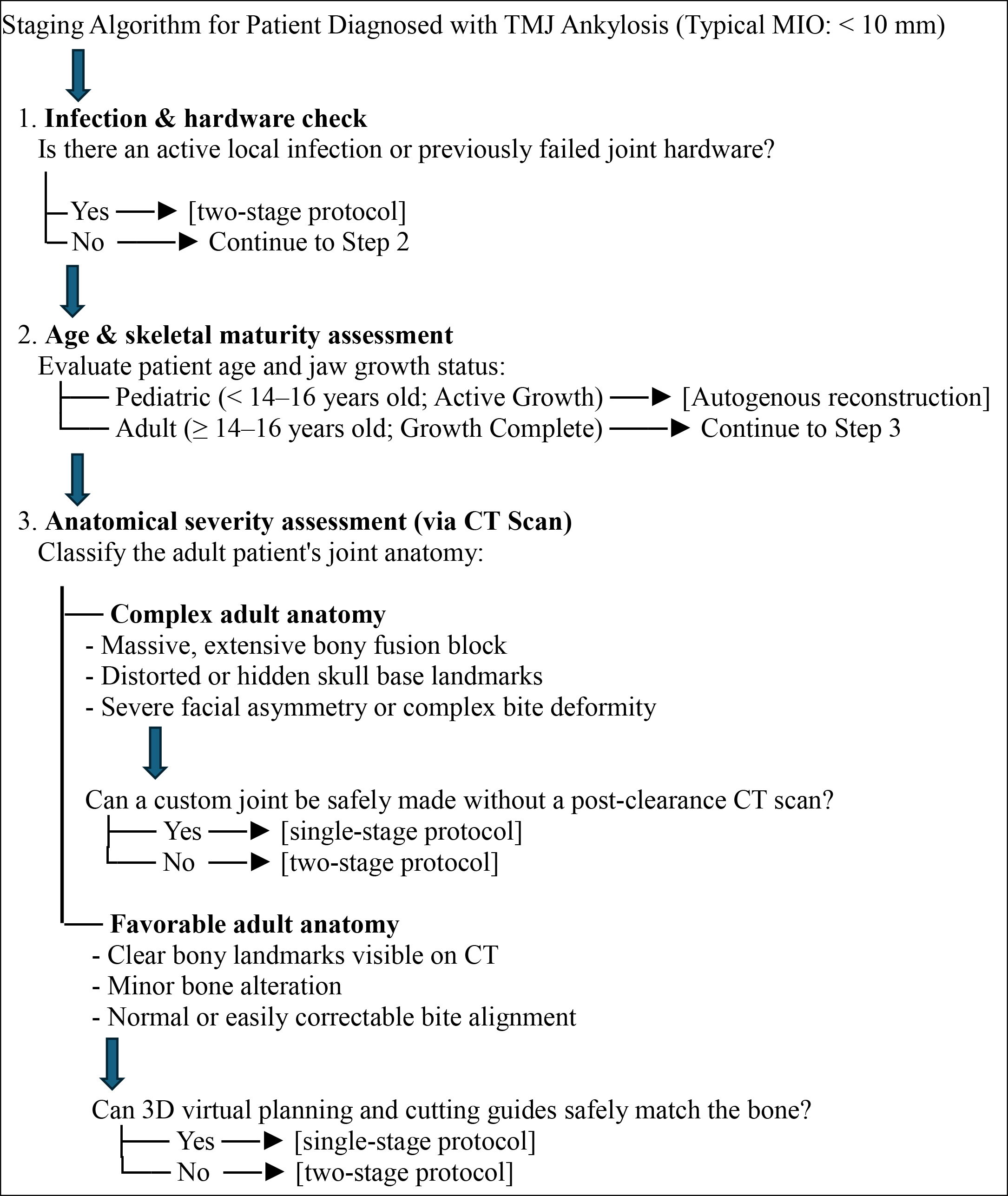
The two-stage protocol involves osteoarthrectomy of the bony ankylosis and the placement of an inert spacer ( Fig. 3 ). A delayed secondary procedure is performed months later to reconstruct the joint with an alloplastic or autogenous graft. It is best suited for: (a) Significant bony ankylosis requiring extensive resection (b) Fossa or condyle anatomy needing major modification (c) Large tumor resections with associated hard-tissue defects (d) Removal of previously failed alloplastic hardware and, (d) Cases requiring significant occlusal changes. This has the advantage of confirmation of no reankylosis before placing the final prosthesis and provides time for presurgical orthodontic preparation, if orthognathic surgery is planned with joint replacement ( Boxes 2 and 3 ).
Intraoperative photograph of a patient showing silastic spacer fixed to temporal bone using wire as a temporary measure for Stage II definitive reconstruction.
Box 2
Box showing 2-stage protocol for reconstruction in temporomandibular joint ankylosis
-
Two-stage protocol (complex adult cases or revision cases)
-
Sequence:
-
First Surgery → Interim Stage (evaluation of reankylosis → Second Surgery → Recovery and Patient-specific implant (PSI) Manufacturing)
-
-
Stage Details:
-
•
Step 1: First Surgery: Osteoarthrectomy. Place a temporary PMMA bone cement spacer or medical grade silastic to hold the space open and keep jaw stable.
-
•
Step 2: Interim Phase (6–12 weeks):
-
Use new CT scan to design the final custom joint.
-
Perform any needed intermediate orthodontics.
-
-
•
Step 3: Second Surgery: Reopen site. Remove the temporary spacer. Clear new scar tissue. Perform replacement.
-
•
Step 4: Recovery: Start intensive physical therapy.
-
•
-
Targets:
-
•
Patient Age: ≥14 to 16 year old
-
•
Stage 1 On table MIO Target: greater than 30 mm intermediate clearance gap
-
•
Postoperative MIO Target: 35 mm to 42 mm achieved after final reconstruction
-
•
Box 3
Staging algorithm for patient diagnosed with temporomandibular joint ankylosis
Osteoarthrectomy planning
-
•
Preoperative CT scans are carefully evaluated, and the full extent of ankylosis is delineated and marked.
-
•
The superior osteotomy is planned just inferior to the zygomatic arch, extending medially up to the full medial extent of the ankylotic mass. Any rudimentary or deformed condylar remnant, if present, should also be identified and excised.
-
•
The inferior osteotomy is planned approximately 1 to 1.5 cm below the superior cut, or as dictated by the planned fossa component in cases where TJR is contemplated.
-
•
If TJR is planned, the safe distance of osteotomy from middle cranial fossa (8 mm), external auditory canal (6–8 mm), and inferior osteotomy to lingula (3–4 mm) is measured.
-
•
In recurrent cases, a temporal bone breach may be present due to previous surgical intervention. If a defect is identified on CT imaging, a preoperative MRI should be obtained to rule out encephalocele and to facilitate appropriate planning—either for extended TJR alone or for a combined neurosurgical and maxillofacial surgical approach.

Stay updated, free dental videos. Join our Telegram channel

VIDEdental - Online dental courses


