Case• 10. A lump on the gingiva
SUMMARY
A 48-year-old man presents to you in general dental practice with a gingival swelling. What is the cause and what would you do?
History
Complaint
The patient complains of a lump on the gum at the front of his mouth on the left side. It sometimes bleeds, usually after brushing or eating hard food but it is not painful.
History of complaint
The swelling has been present for 4 months and has grown slowly during this period. It was never painful but now looks unsightly. The patient gives no history of other mucosal or skin lesions.
Medical history
The patient has hypertension, controlled with atenolol 50 mg daily.
Examination
Extraoral examination
He is healthy looking but slightly overweight. There are no palpable cervical lymph nodes.
Intraoral examination
The patient is partially dentate and has relatively few and extensively restored teeth. He wears an upper partial denture. The root of the upper lateral incisor is present and its carious surface lies at the level of the alveolar ridge. The teeth on each side of the lesion are restored with metal–ceramic crowns.
There is a mild degree of marginal gingivitis. Most of the interdental papillae are rounded and marginal inflammation is present around crowns. Flecks of subgingival calculus are visible.
▪ The appearance of the lesion is shown inFigure 10.1. Describe its features.
| Feature | Appearance |
|---|---|
| Site | Appears to arise from the gingival margin of the lateral incisor root or the interdental papilla mesially |
| Size | Approximately 10 × 7 mm |
| Shape and contour | Irregular rounded nodule. It is not possible to say whether it is pedunculated or sessile, though from its size and the fact that it overlies the lateral incisor root, it is probably pedunculated |
| Colour | Patchy red and pink with a thin grey translucent sheen. The surface is almost certainly ulcerated |
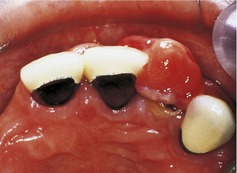 |
| Fig. 10.1 |
If you were able to palpate the lesion you would find that it is fleshy and soft and attached by a thin base to the gingival margin. It bleeds readily from between the tooth and lesion when pressed with an instrument but it is not tender.
▪ From the information in the history and examination so far, what is your differential diagnosis?
Likely:
— Pyogenic granuloma (if the patient had been female, pregnancy epulis might have been considered)
— Fibrous epulis
Less likely:
— Peripheral giant cell granuloma
— Sinus papilla (parulis)
Unlikely:
— Papilloma
— Benign hamartoma or neoplasm
— Malignant neoplasm.
▪ Justify your differential diagnosis.
A very wide range of lesions may affect the gingiva and many possible causes cannot be excluded on the basis of the information given so far. However, the gingiva is the site of predilection for a number of inflammatory hyperplastic lesions comprising fibrous tissue and epithelium. All are associated with poor oral hygiene and the lesion is almost certainly one of this type on statistical grounds.
Pyogenic granuloma is a localized proliferation of granulation tissue or very vascular fibrous tissue. It arises in association with a local irritant such as poor oral hygiene, calculus or the margin of a restoration. The present lesion has many features of the pyogenic granuloma: it is asymptomatic, soft and vascular, bleeds readily, and has an ulcerated surface. If the patient had been female, a pregnancy epulis (a variant of pyogenic granuloma arising during pregnancy) would have been possible.
Stay updated, free dental videos. Join our Telegram channel
VIDEdental - Online dental courses


