Case• 53. Unexpected findings
SUMMARY
A 14-year-old boy presents with toothache and a slightly swollen left cheek. What is the diagnosis and how will you treat him?
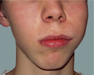 |
| Fig. 53.1. |
| The patient’s appearance on presentation. |
History
Complaint
The patient complains of intermittent toothache on the left side of his face, which he feels is coming from an upper tooth.
History of complaint
He has been aware of intermittent pain and discomfort from an upper back tooth when eating, especially anything very hot or cold, for several months. The pain is gradually getting worse.
Medical history
The patient is otherwise fit and well.
Dental history
He has never been to a dentist before.
Examination
Extraoral examination
The patient is a fit and healthy looking boy. His left cheek appears slightly swollen but there is little extraoral asymmetry. The cheek is not tender or inflamed and both the patient and his parents say that he has always looked like this. No lymph nodes are palpable and his temporomandibular joints appear normal.
Intraoral examination
The upper left first molar has heavily stained fissures and the whole crown is discoloured. The other teeth appear sound. The alveolus in the upper left quadrant is enlarged, with reduction of depth of the buccal sulcus. The swelling affects the buccal and palatal aspects, is smooth, uninflamed and is not tender to palpation.
In addition, the upper left second premolar appears missing and there is a small space between the first premolar and molar tooth. Several supernumerary teeth are evident.
▪ How do you interpret the history and examination so far?
There could be several explanations for the presentation. The history of the pain, exacerbated by hot and cold, and poorly localized almost certainly indicates pulpitis. The obvious cause would appear to be caries in the upper first molar. The whole crown is discoloured and there may be extensive caries despite the intact occlusal surface.
The enlargement of the alveolus has expanded into the buccal sulcus and could account for the slight extraoral swelling. The commonest cause of smooth uninflamed expansion of the alveolus is an odontogenic cyst. Further investigation is required.
The absent second premolar may be unerupted or absent. Missing premolar teeth is a relatively common developmental anomaly but the patient also has supernumerary teeth and it would be unusual to have missing and supernumerary teeth in the same patient. In addition, if the premolar had never developed, the space between the first premolar and molar would have been likely to have closed completely. The tooth is probably unerupted and relatively superficial in the alveolus, holding the teeth apart.
Investigations
▪ What investigations would you now undertake and why?
Vitality tests. The vitality of the upper first molar needs to be determined and on testing it you discover that it appears vital, as are the adjacent teeth.
Radiographs. Right and left bitewings for caries assessment and a panoramic radiograph to assess the overall dentition are indicated. These views should provide sufficient information to explain the missing upper left second premolar, assess any further unerupted supernumerary teeth and to investigate the swelling of the left maxilla. If required, further views including periapicals, oblique upper occlusal views or antral views may be required later but are not indicated at this stage.
▪ The panoramic radiograph is shown inFigure 53.2. Look carefully. What do you see?
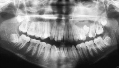 |
| Fig. 53.2 |
The panoramic radiograph shows:
• a large carious cavity in upper first molar
• right and left supplemental maxillary canines
• peg-shaped supernumerary overlying upper right lateral incisor and right canine
• upper left second premolar present, unerupted and inverted
• developing third molars in all four quadrants
• increased opacity in the region of the left maxillary sinus.
▪ What terms are used to describe extra teeth? What do they mean?
| Term | Definition |
|---|---|
| Supernumerary tooth | Any tooth over and above the normal complement of teeth. |
| Supplemental tooth | Supernumerary tooth with the morphology of a normal tooth, usually an additional tooth in a series, for instance additional lateral incisor, third premolar or fourth molar. |
| Mesiodens | Supernumerary tooth in the upper midline, may be conical (forms early and rarel/> |
Stay updated, free dental videos. Join our Telegram channel
VIDEdental - Online dental courses


