Case• 39. Fractured incisors
SUMMARY
A 38-year-old man presents to you in your local hospital accident and emergency department. He has fractured his front teeth. You must manage the injury and outline a treatment plan for restoration.
History
Complaint
The patient’s front teeth have been fractured and they are all loose. One tooth was knocked out and he feels pain when he bites.
History of complaint
The car accident occurred yesterday. The patient was sitting in the driver’s seat when another car drove into his. He was stationary and not wearing a seat belt and was thrown forward, his lower face hitting the steering wheel. He did not lose consciousness and was taken to a local accident and emergency department where a laceration of his lower lip was sutured and no other injuries were found. At that time his teeth and jaws were not examined or radiographed and he has returned for a follow up appointment with you.
Medical history
Prior to the accident the patient was fit and healthy, with only allergy to penicillins and erythromycin noted on his medical history questionnaire.
Examination
Extraoral examination
▪ How will you assess the possibility of a mandibular fracture?
Fracture is suggested by:
• pain, swelling and tenderness at the fracture site
• bleeding, bruising or haematoma at fracture site
• displacement, step deformity
• change in occlusion
• mobility of fragments or of teeth
• difficulty opening the mouth or in lateral excursion
• paraesthesia or anaesthesia in the distribution of nerves involved in the fracture.
▪ How will you assess the possibility of a fracture of the zygomatic arch or facial skeleton?
In addition to the features noted above, fracture at these sites may produce:
• facial asymmetry and flattening of facial contour (may be masked by swelling for a few days)
• step deformity along infraorbital margin
• anaesthesia or paraesthesia of cheek, nose, upper lip and teeth
• unilateral epistaxis
• subconjunctival haemorrhage with no definable posterior limit
• restricted eye movements and diplopia.
On extraoral examination you cannot identify a mandibular or facial fracture. The lower lip is swollen and lacerated to the left of the midline. There is no restriction or pain on opening, nor swelling associated with the temporomandibular joint.
Intraoral examination
▪ The anterior teeth are shown inFigure 39.1. What do you see?
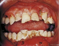
The swollen lower lip is just visible. The upper right lateral and both central incisors have been fractured. The upper left lateral incisor appears to be missing. The upper left canine is not fractured but has caries buccally and is mesially inclined. This inclination could predate the injury, in which case the lateral incisor may have been buccally positioned, or it could be a result of injury.
The oral hygiene is poor. This does not appear to be a result of injury because there are large accumulations of plaque, staining of the teeth, inflammation of the gingiva and caries buccally on teeth in the upper arch.
▪ What investigations would you ensure had been carried out at the patient’s first attendance? Explain why.
A posterior–anterior radiograph of the chest should have been taken to determine whether the lost tooth or any fragments were inhaled.
A soft tissue radiograph of the lower lip in the region of the laceration should have been taken before suturing. This would exclude the possibility that small tooth fragments have been embedded in the lip. Two views at right angles would be required to localize small fragments and surgical removal can be facilitated if radiopaque markers are taped to the skin before radiography. These markers may be used to localize the fragment at removal. Larger fragments of tooth in the lip are usually obvious on examination and debridement of the wound. Such fragments may cause infection or become embedded in scar tissue, sometimes deforming the lip.
Facial radiographs should have been taken to assess the possibility of an undisplaced facial fracture of the maxilla, zygoma or mandible. A suitable selection of films for this purpose would be a 10° and 30° occipitomental view, posterior–anterior jaws and a dental panoramic radiograph.
These films may have been overlooked at first examination if the fractured teeth were not recognized or if casualty staff concentrated on excluding more serious injuries. It is obviously undesirable for the patient to undergo radiography again.
Stay updated, free dental videos. Join our Telegram channel
VIDEdental - Online dental courses


