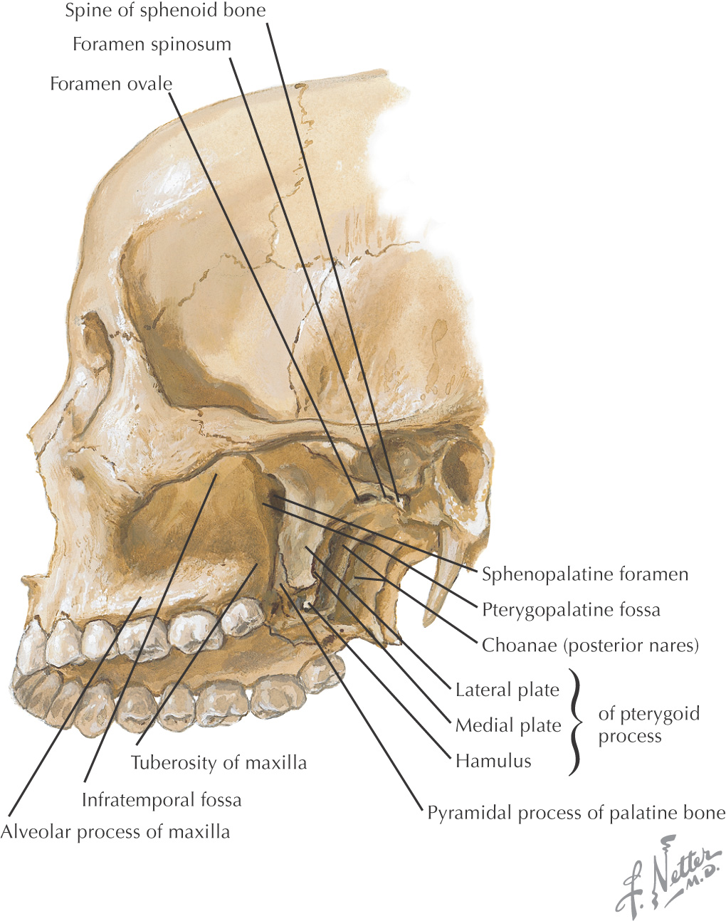
PTERYGOPALATINE FOSSA
Overview and Topographic Anatomy
Contents of the Pterygopalatine Fossa
Overview and Topographic Anatomy
GENERAL INFORMATION
Pyramid-shaped fossa on the lateral aspect of the skull between the maxilla’s infratemporal surface and the pterygoid process of the sphenoid
Contains major nerves and blood vessels that supply the nasal cavity, upper jaw, hard palate, and soft palate: the maxillary division of the trigeminal nerve, pterygopalatine (sphenopalatine, Meckel’s) ganglion, and 3rd portion of the maxillary artery
Allows the infratemporal fossa, middle cranial fossa, foramen lacerum, nasopharynx, nasal cavity, orbital cavity, and oral cavity to communicate
7 foramina/fissures allow passage of nerves and vessels
Borders and Openings
BORDERS
|
Border |
/> |
Stay updated, free dental videos. Join our Telegram channel
VIDEdental - Online dental courses


