15: Pharynx

CHAPTER 15

PHARYNX

Overview and Topographic Anatomy

Parts of the Pharynx

Muscles

Potential Apertures in Pharyngeal Wall

Vascular Supply

Nerve Supply

Clinical Correlate

Questions

Overview and Topographic Anatomy

GENERAL INFORMATION

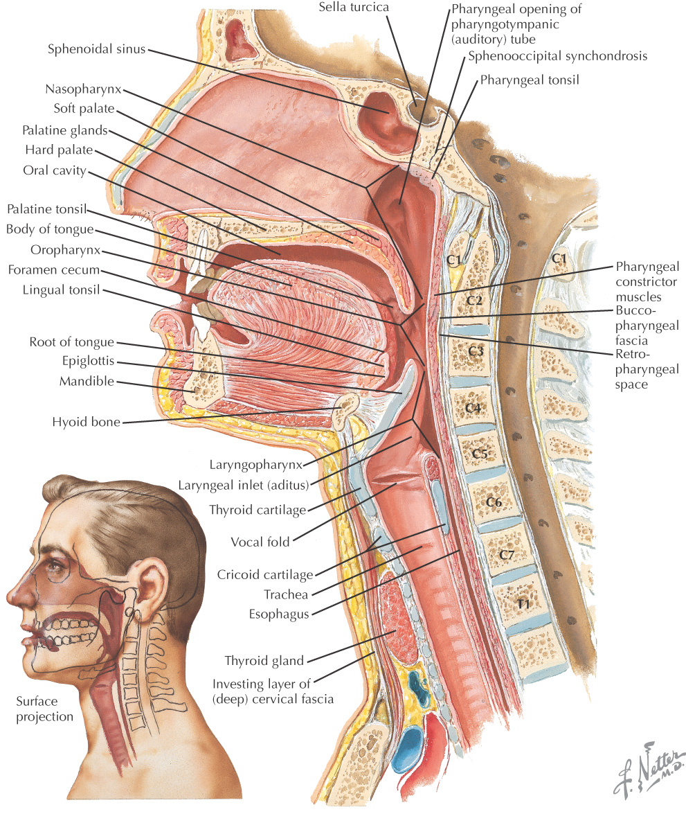
Pharynx: 5-inch muscular tube from base of the skull to the lower border of the cricoid cartilage (C6)

Posterior portion of the pharynx lies against the prevertebral fascia

Lies posterior to the nasal and oral cavities and the larynx and thus is divided into 3 parts:

Nasopharynx

Oropharynx

Laryngopharynx

Responsible for properly conducting food to the esophagus and air to the lungs
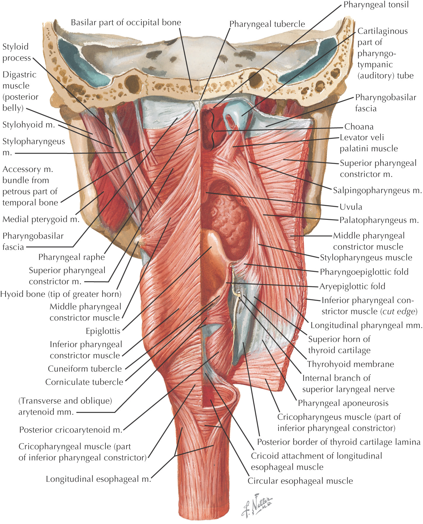
Composed of:

Three constrictor muscles

Three longitudinal muscles

Cartilaginous part of the pharyngotympanic tube

Soft palate

The wall of the pharynx has 5 layers:

Mucous membrane—the innermost layer

Submucosa

Pharyngobasilar fascia—the fibrous layer attached to the skull anchoring the pharynx

Muscular—3 inner longitudinal and 3 outer circular (constrictor) muscles that overlap such that the superior constrictor is the innermost, whereas the inferior constrictor is the outermost muscle

Buccopharyngeal fascia—loose layer of connective tissue continuous with the fascia over the buccinator and pharyngeal muscles, and the location of the pharyngeal plexus of nerves and the pharyngeal plexus of veins

images
images

Parts of the Pharynx

NASOPHARYNX

Only gold members can continue reading. Log In or Register to continue

Stay updated, free dental videos. Join our Telegram channel

Jan 5, 2015 | Posted by in General Dentistry | Comments Off on 15: Pharynx

VIDEdental - Online dental courses

Get VIDEdental app for watching clinical videos