PAROTID BED AND GLAND
Overview and Topographic Anatomy
Overview and Topographic Anatomy
GENERAL INFORMATION
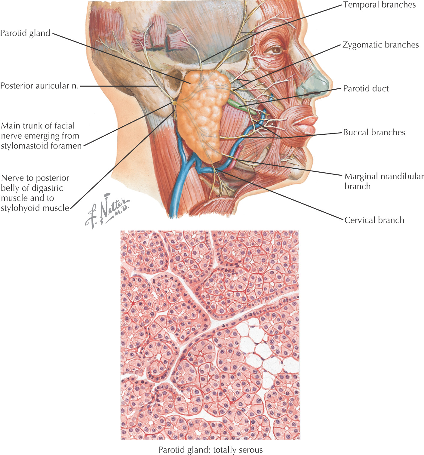
The largest of all the major salivary glands
Entirely serous in secretion
Pyramidal in shape, with up to 5 processes (or extensions)
The gland’s capsule is from the deep cervical fascia
ANATOMIC LANDMARKS
Approximately 75% or more of the parotid gland overlies the masseter muscle; the rest is retromandibular
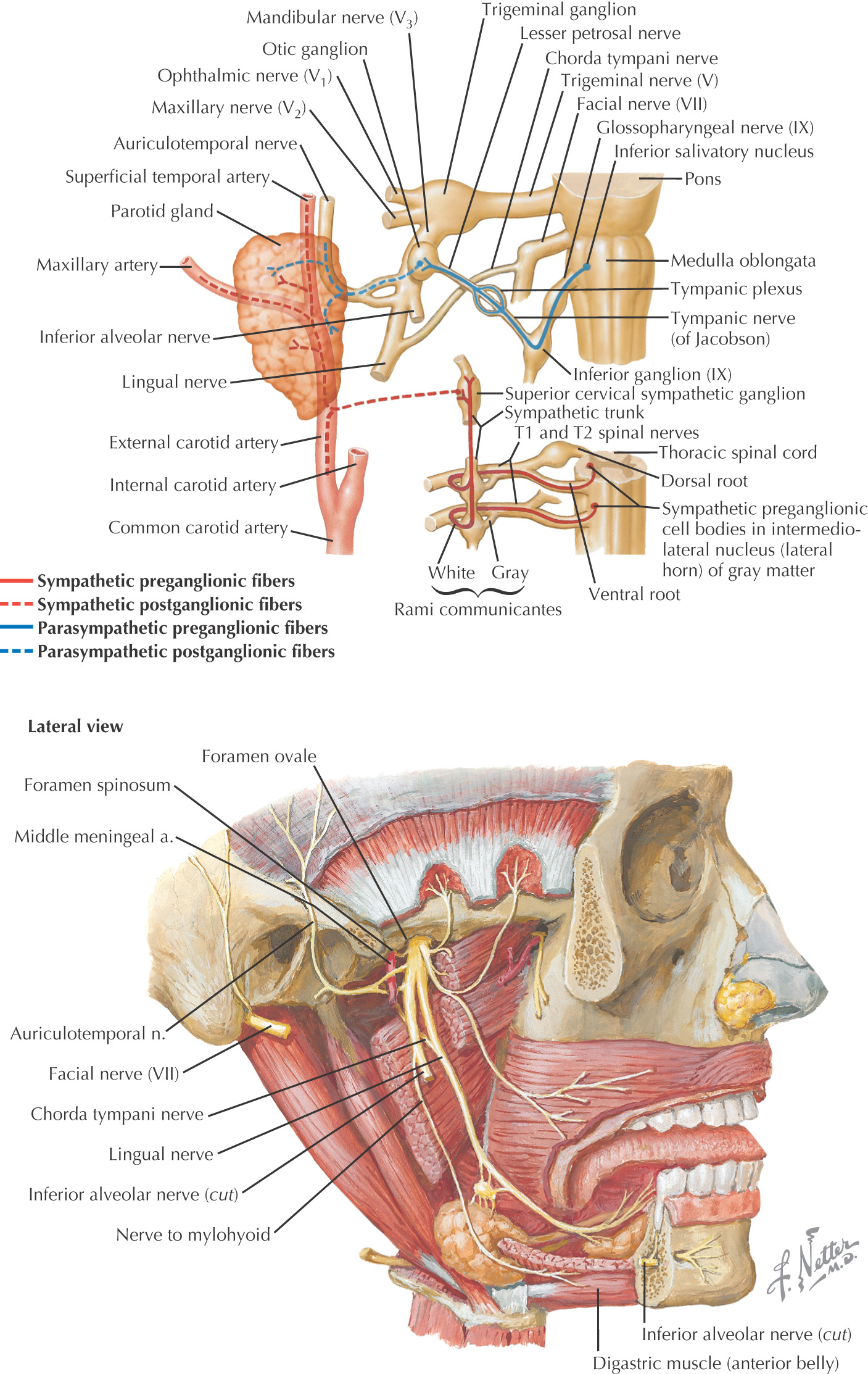
Facial nerve enters the parotid fossa by passing between the stylohyoid muscle and the posterior belly of the digastric muscle, then splits the gland into a superficial lobe and a deep lobe that are connected by an isthmus
Deep lobe lies adjacent to the lateral pharyngeal space
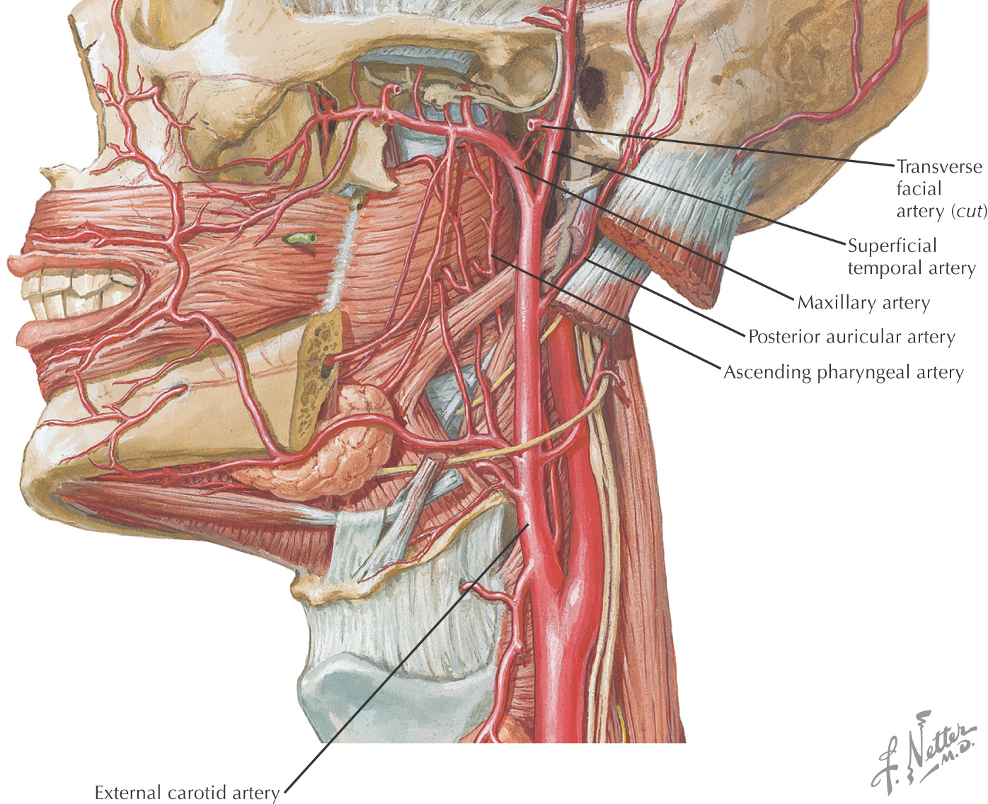
Transverse facial artery parallels the parotid duct slightly superior to the duct
Buccal and zygomatic branches of the facial nerve form an anastomosing loop superficial to the parotid duct
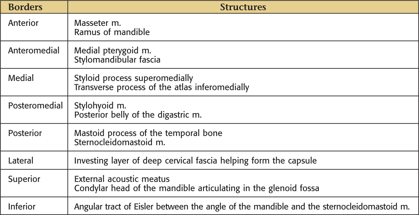
Recess of the Parotid Bed
BORDERS AND STRUCTURES

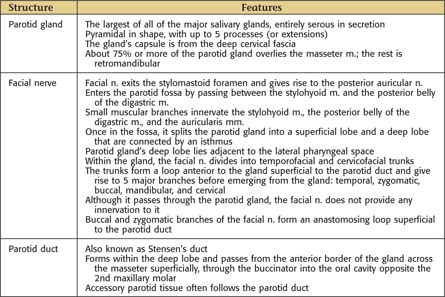
Contents of the Parotid Bed
MAJOR STRUCTURES

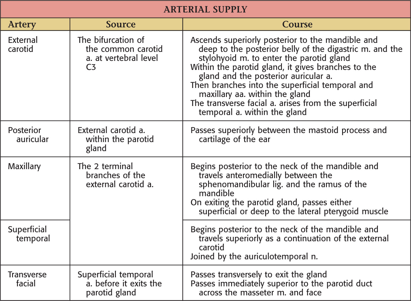
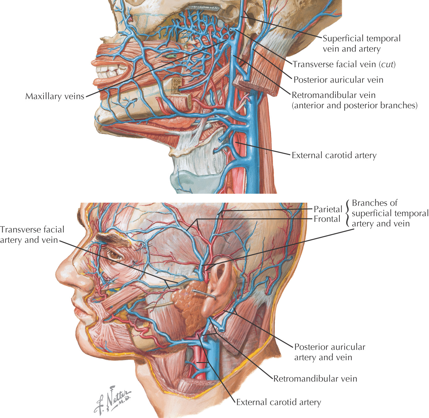
VASCULAR SUPPLY
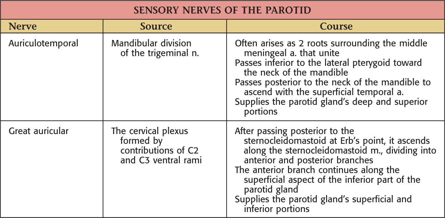
NERVE SUPPLY
Clinical Correlate
BELL’S PALSY
Unilateral facial paralysis from facial nerve (cranial nerve VII) damage
CAUSES
Approximately 80% of cases have unclear etiology
Evidence suggests herpes simplex virus (HSV-1) infection is a cause
•
Stay updated, free dental videos. Join our Telegram channel
VIDEdental - Online dental courses


