INTRODUCTION TO THE UPPER LIMB, BACK, THORAX, AND ABDOMEN
Overview and Topographic Anatomy
Overview and Topographic Anatomy
GENERAL INFORMATION
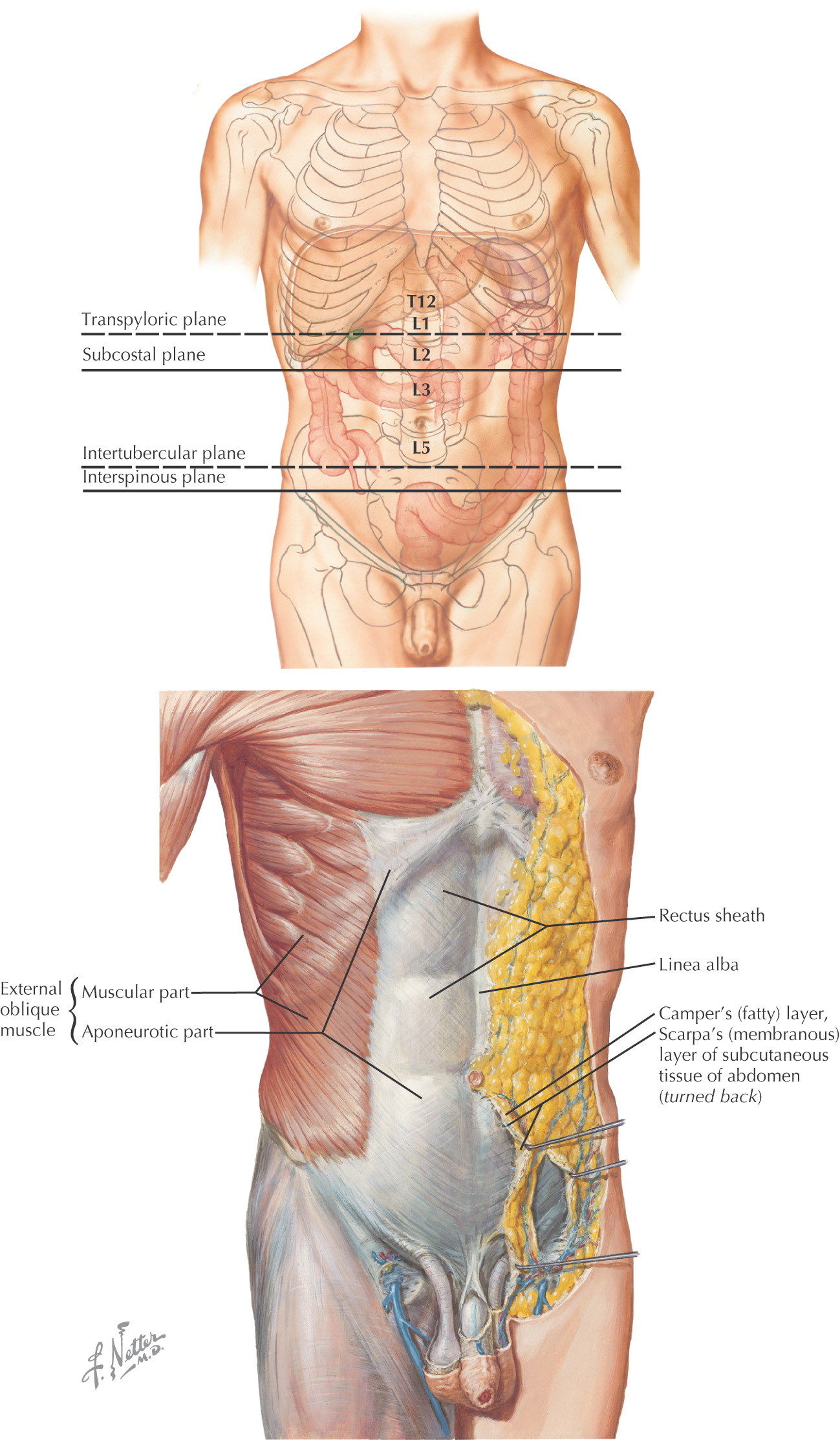
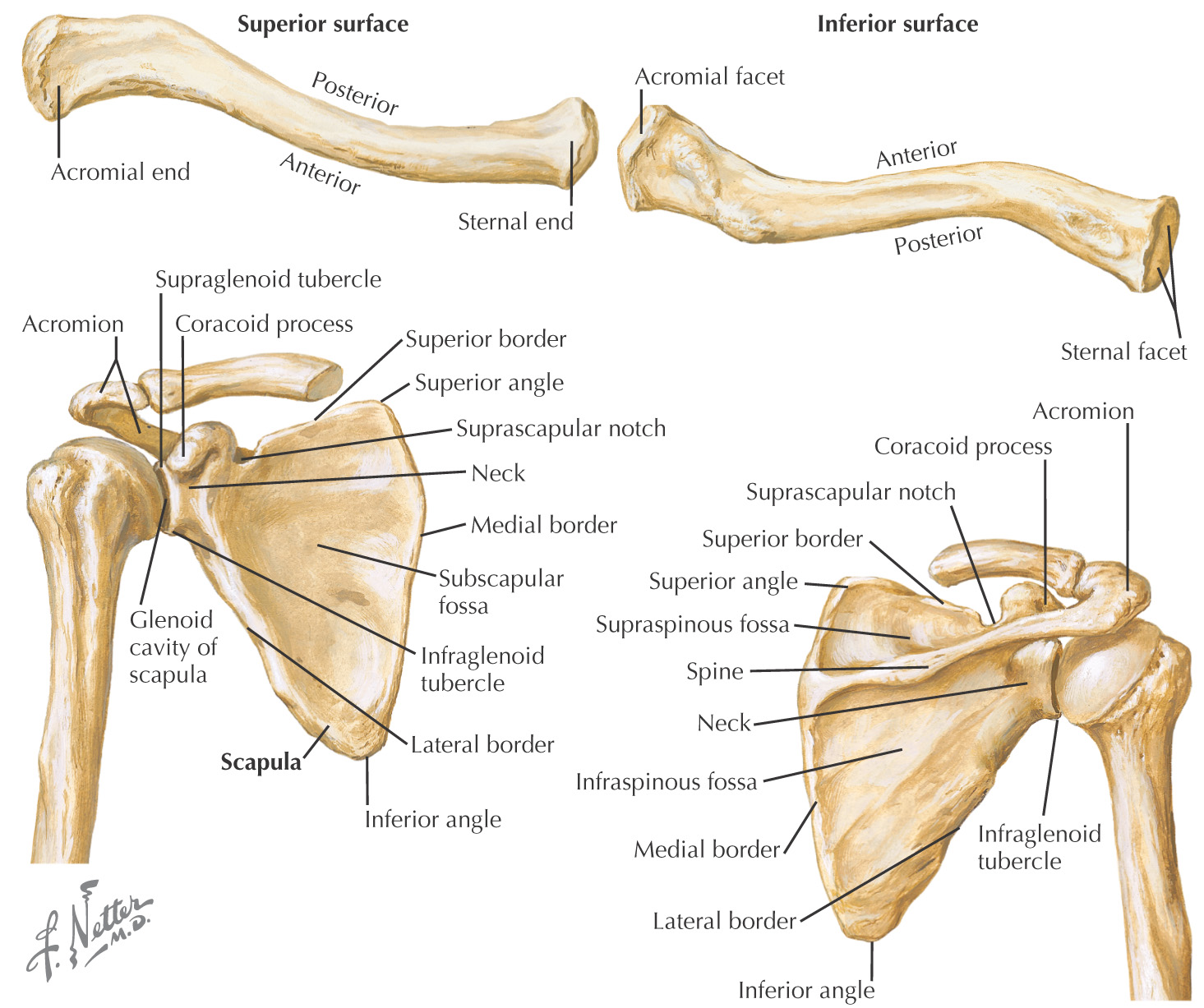
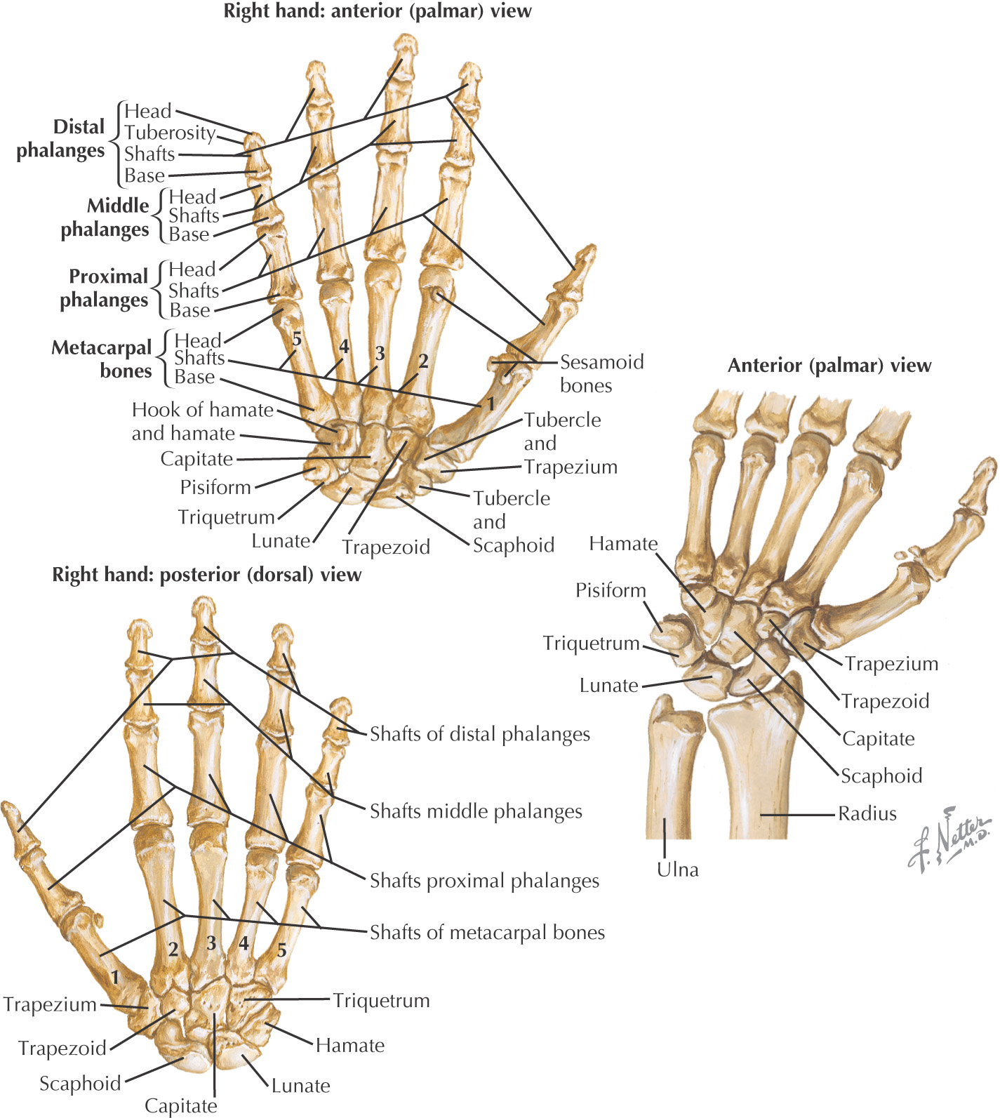
Osteology
UPPER LIMB
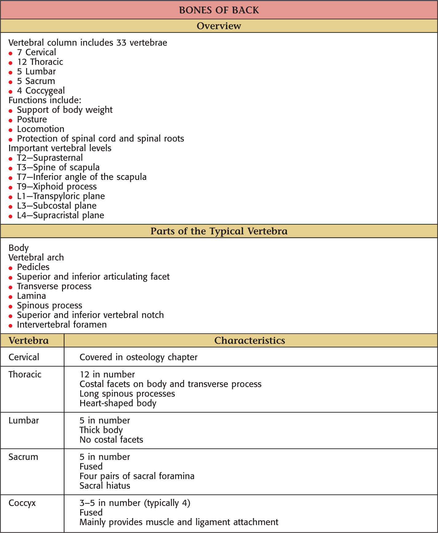
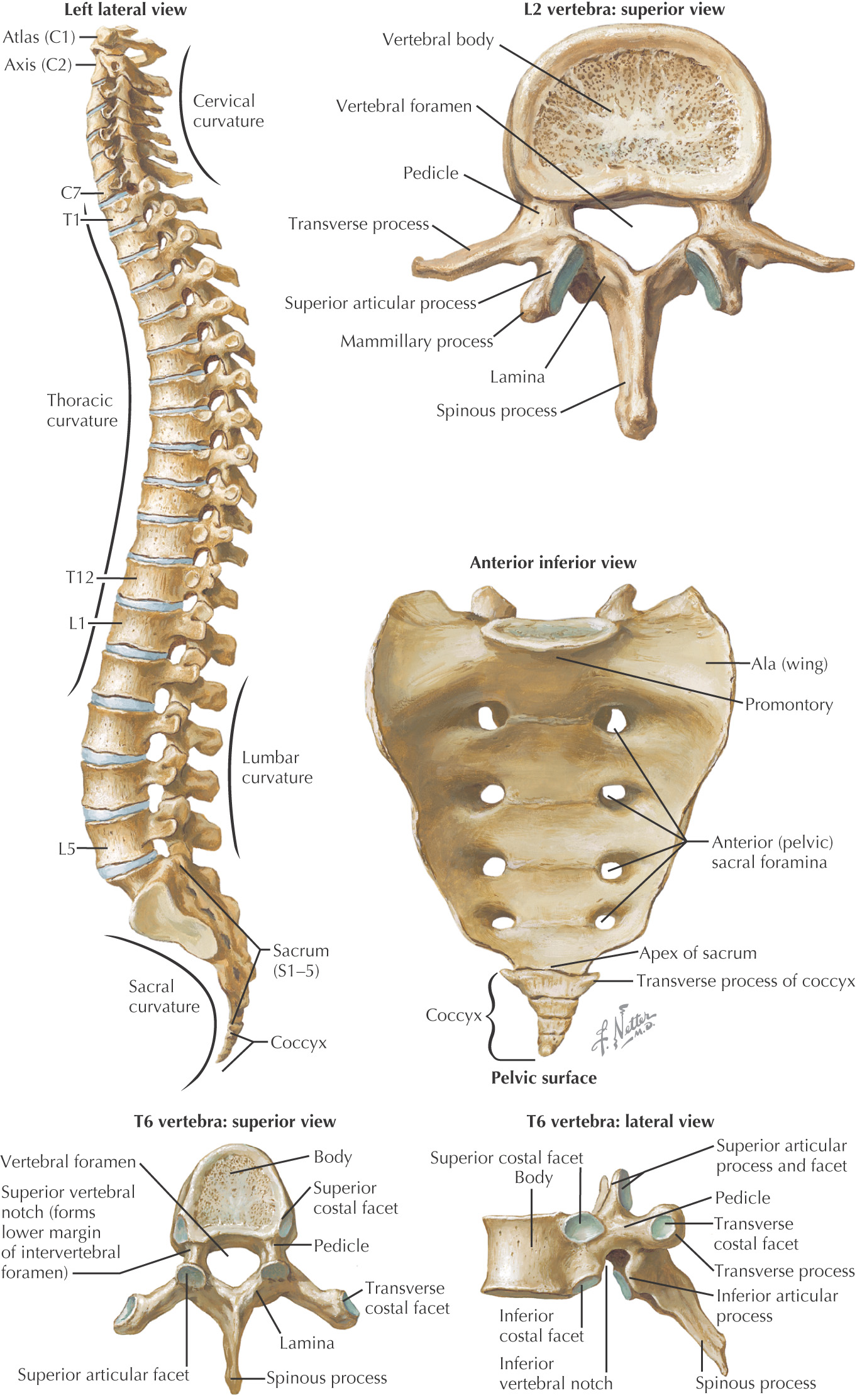
BACK
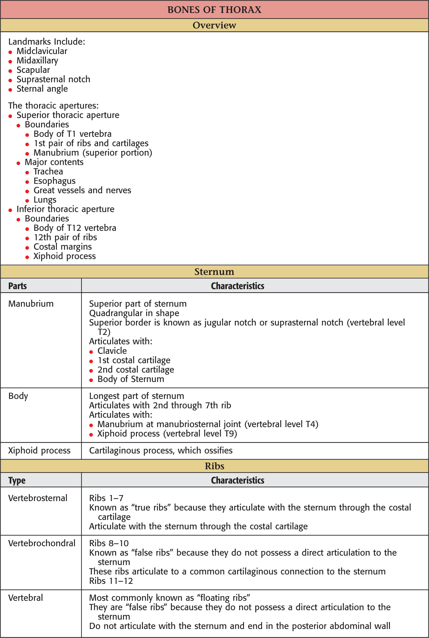
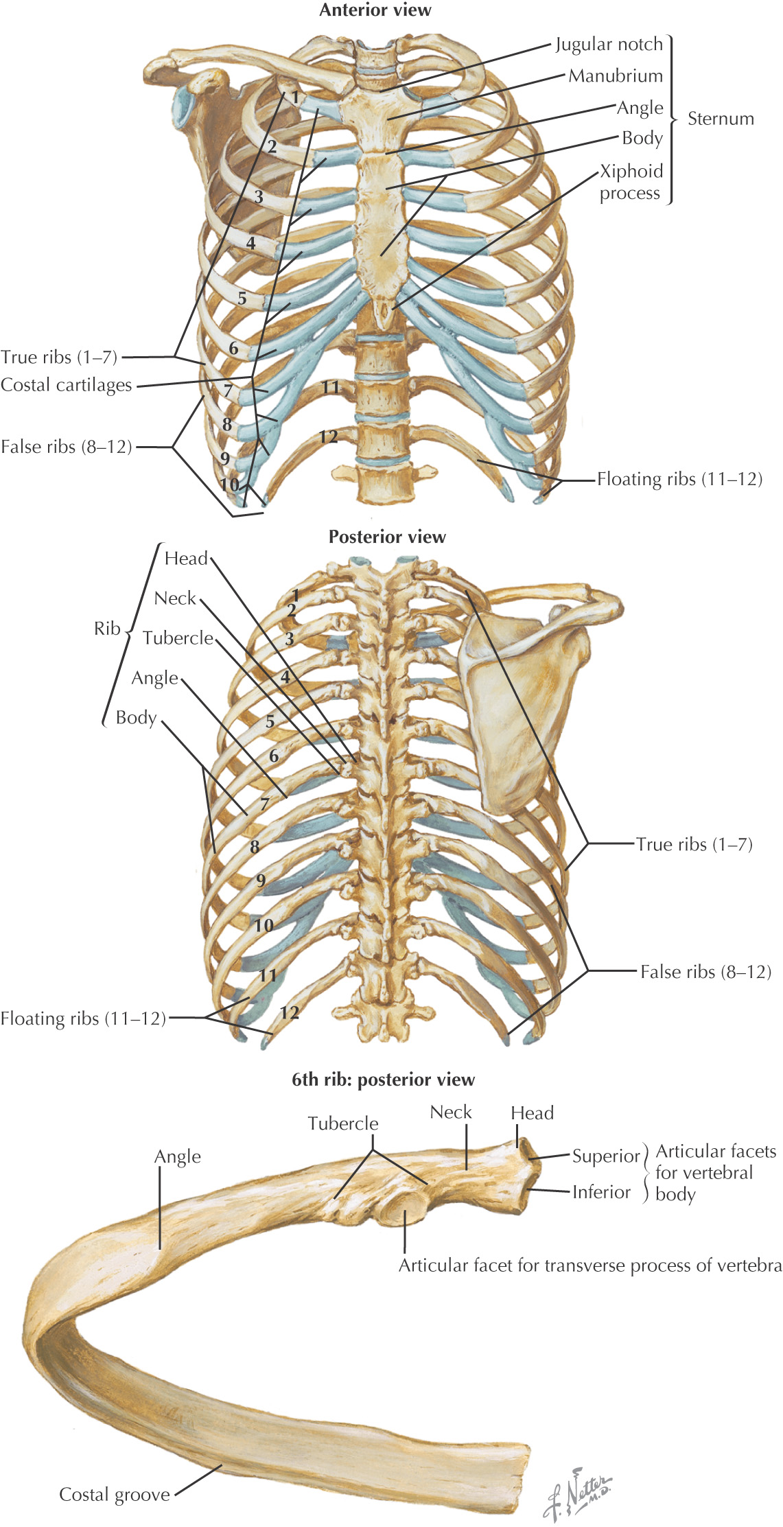
THORAX
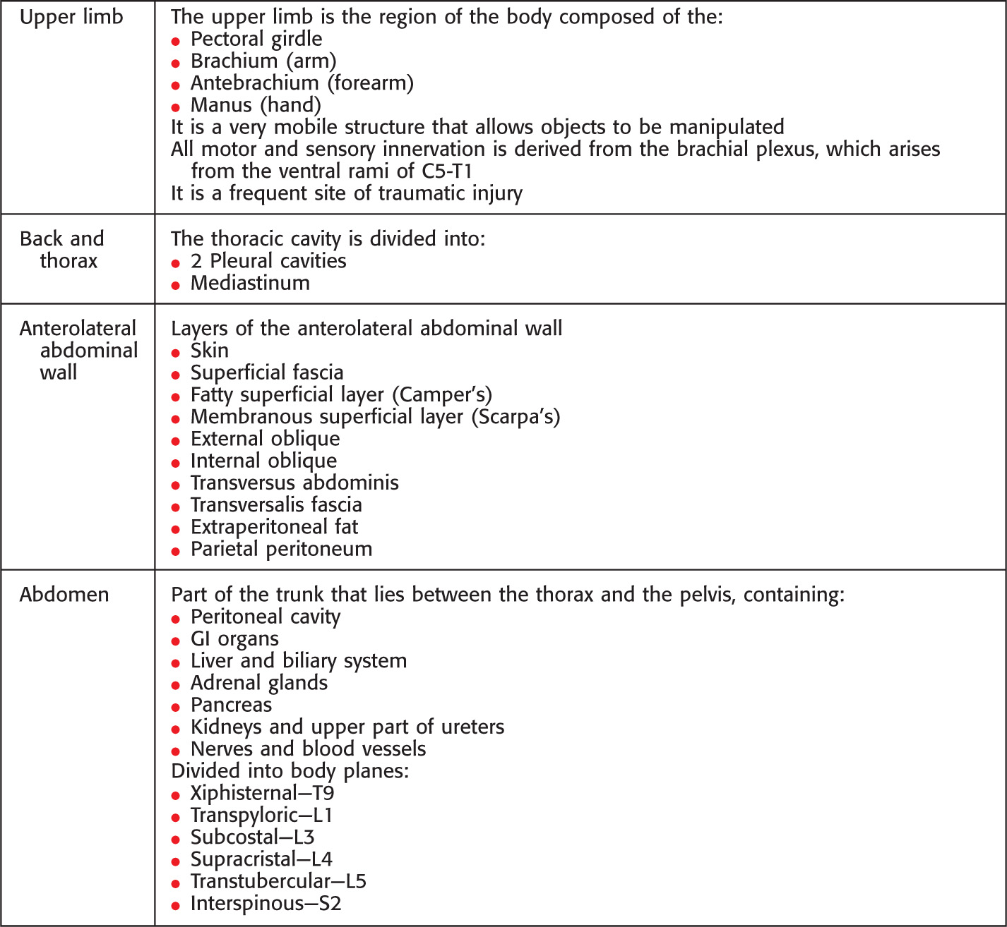
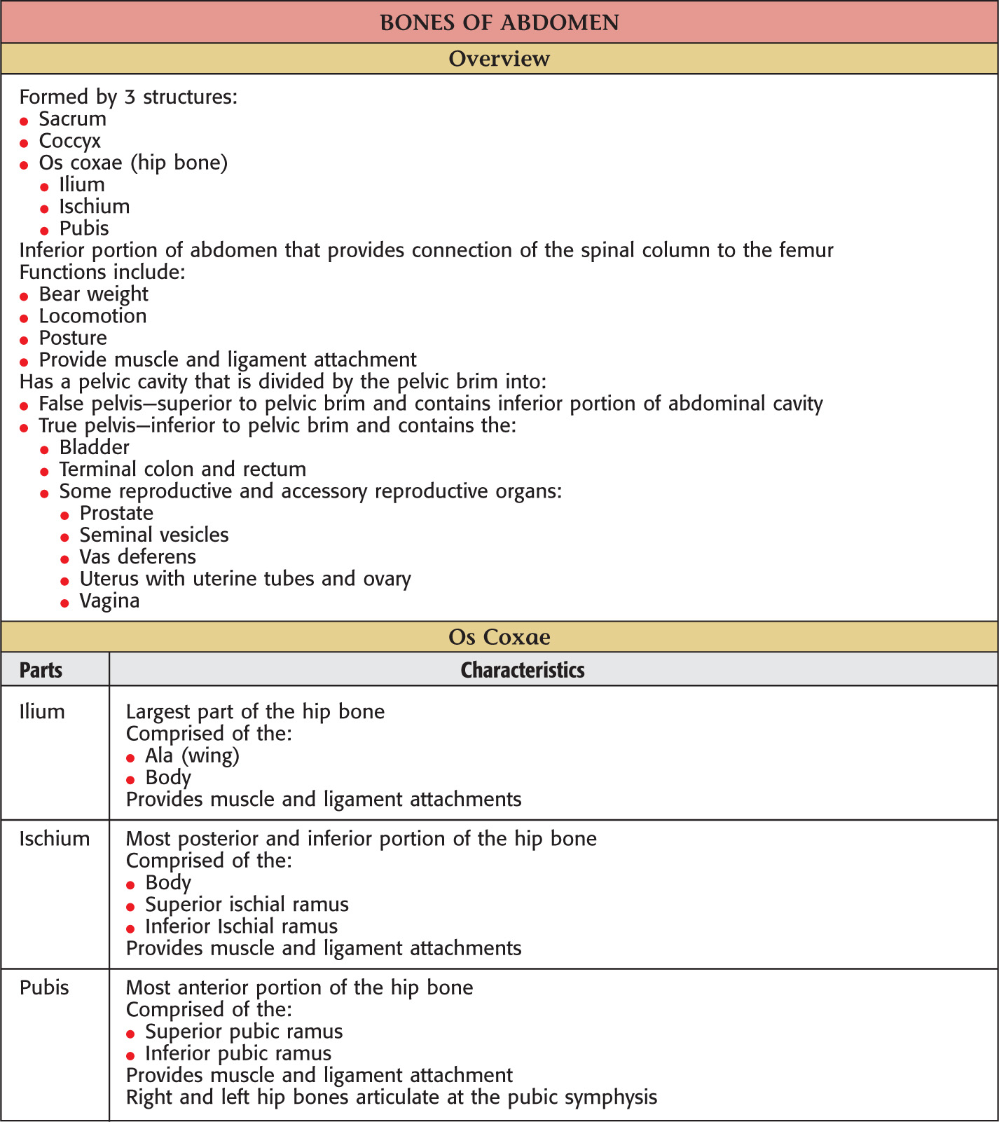
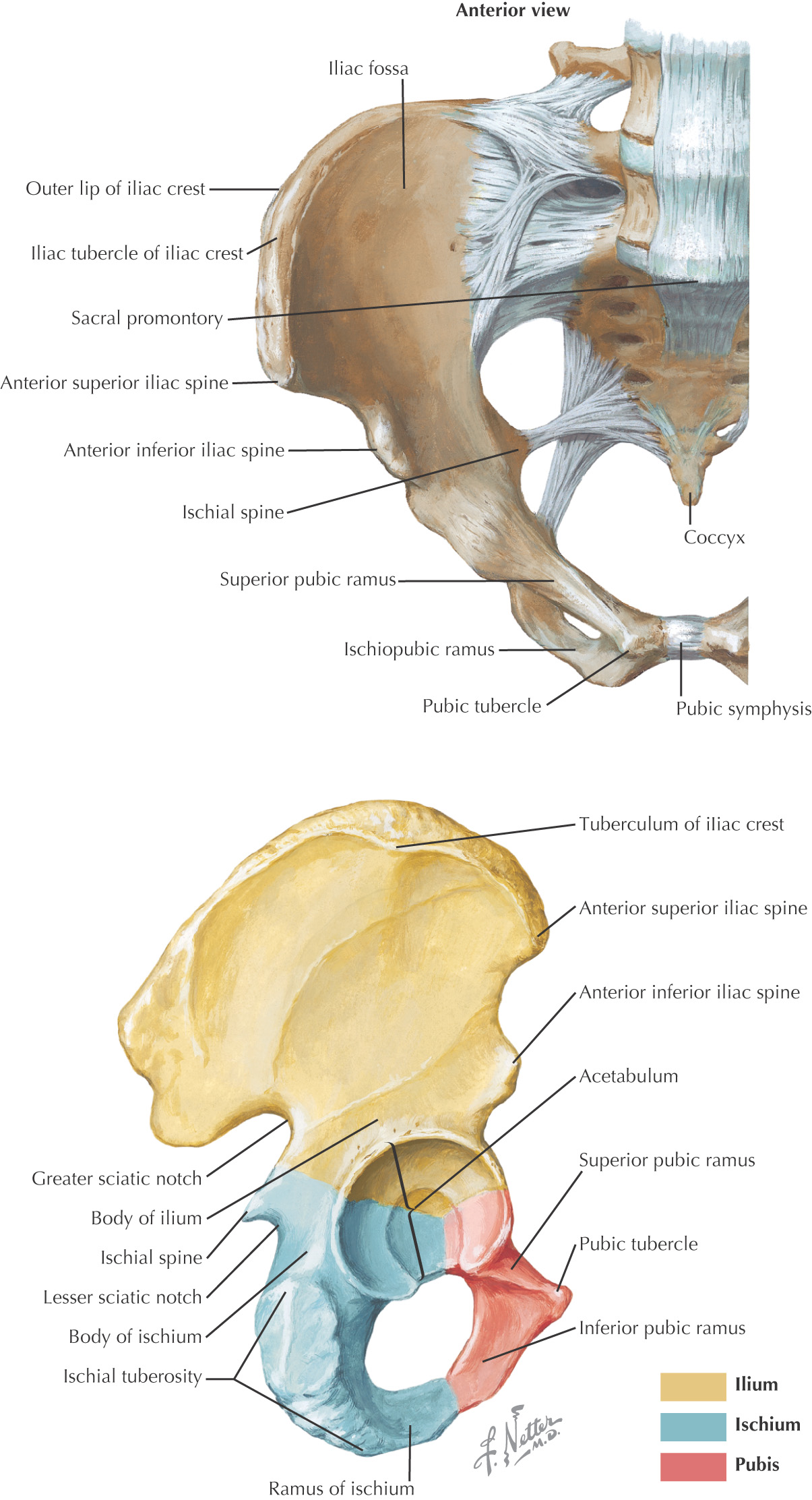
ABDOMEN
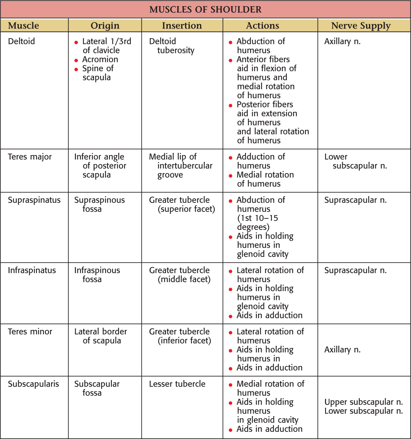
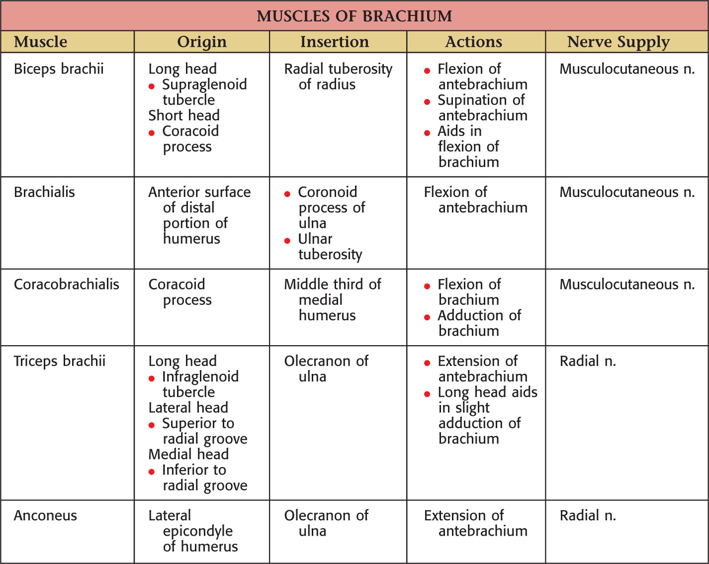
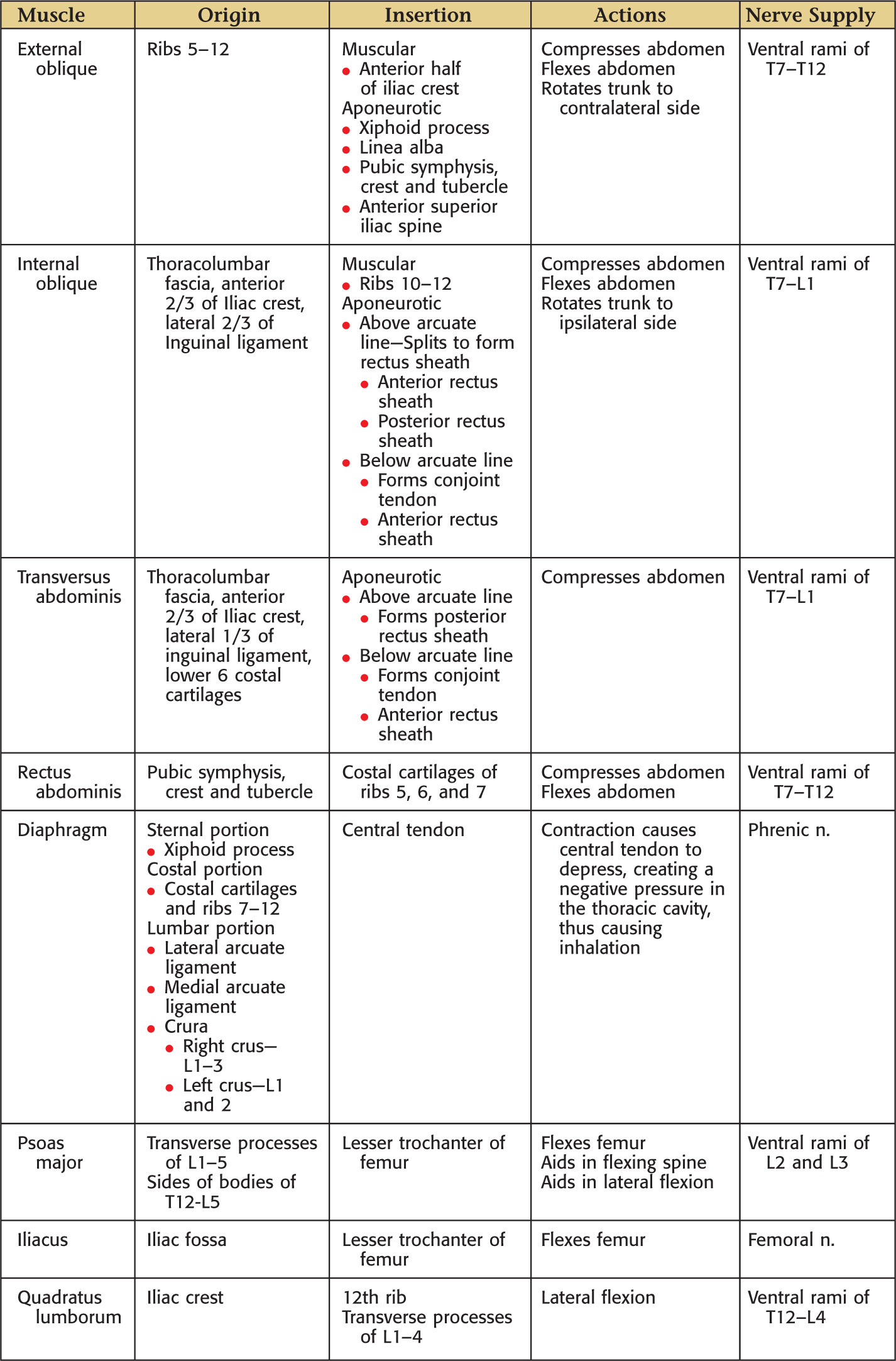
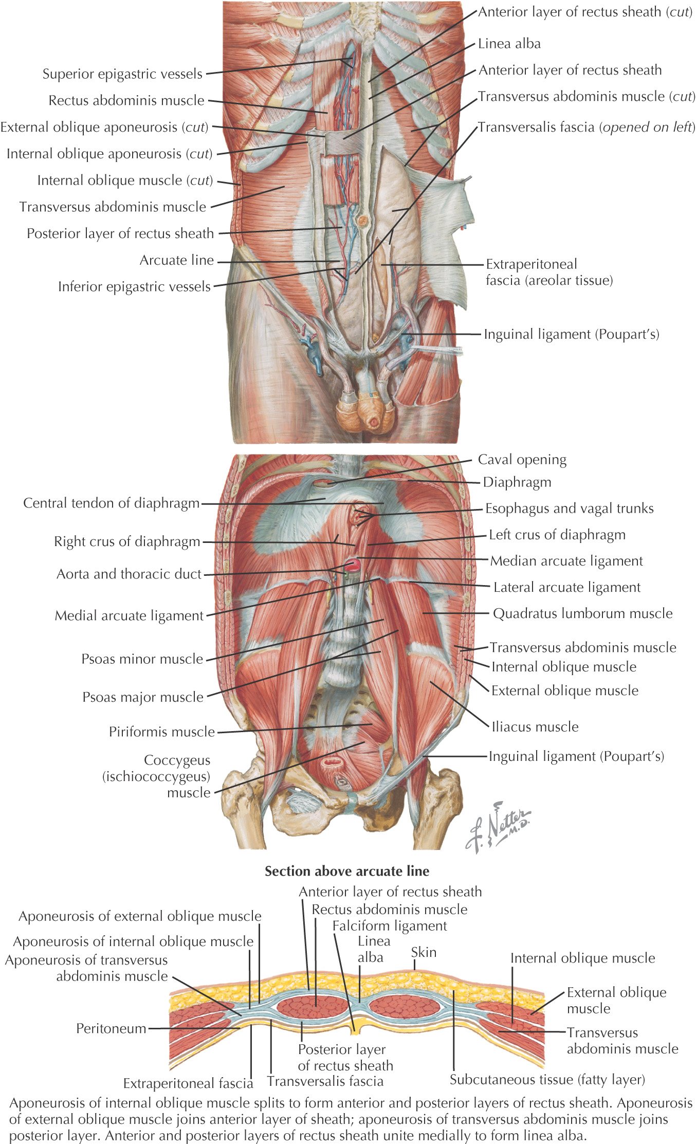
Muscles
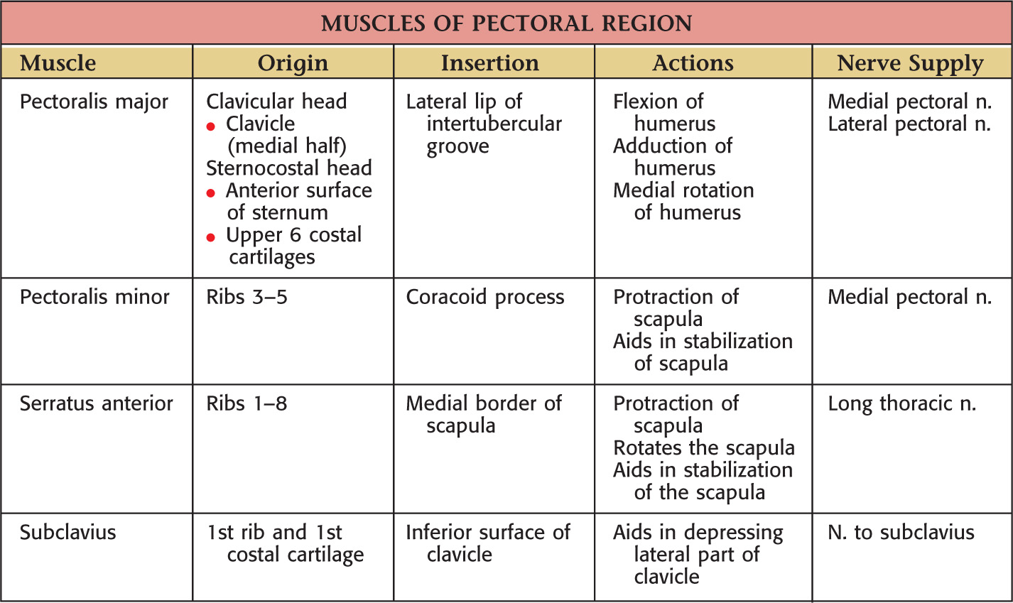
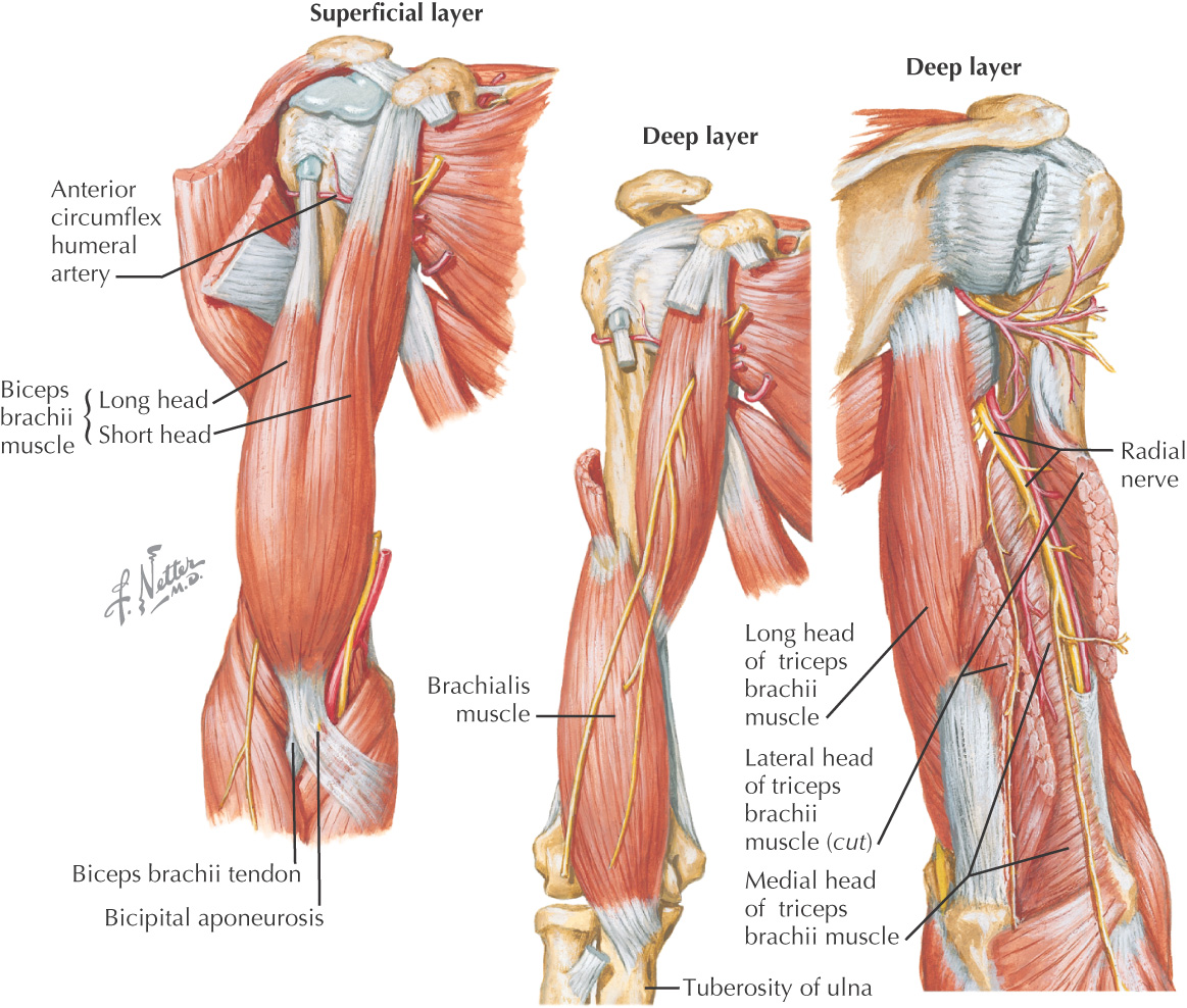
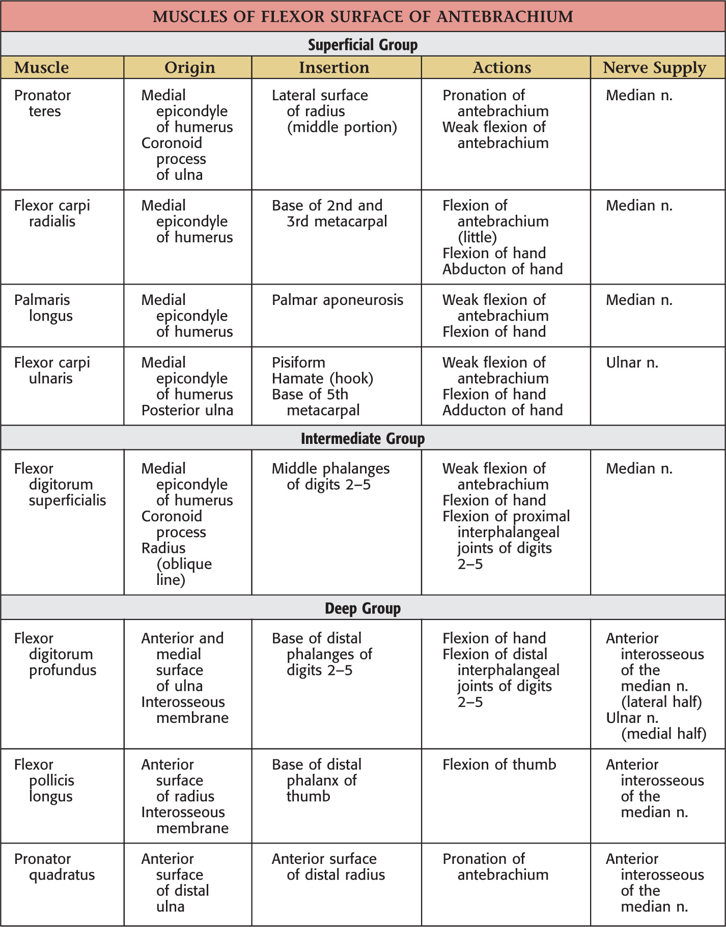
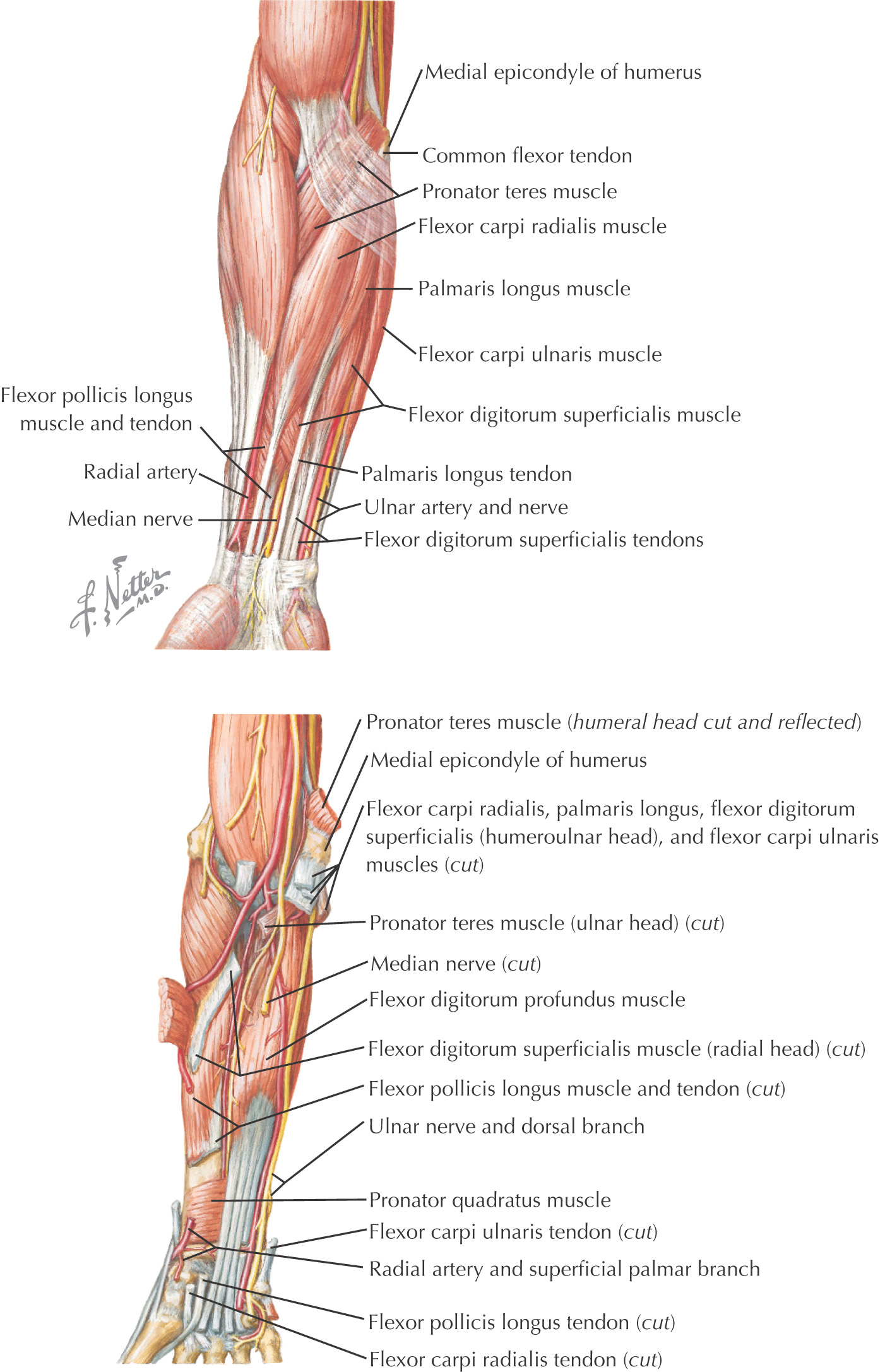
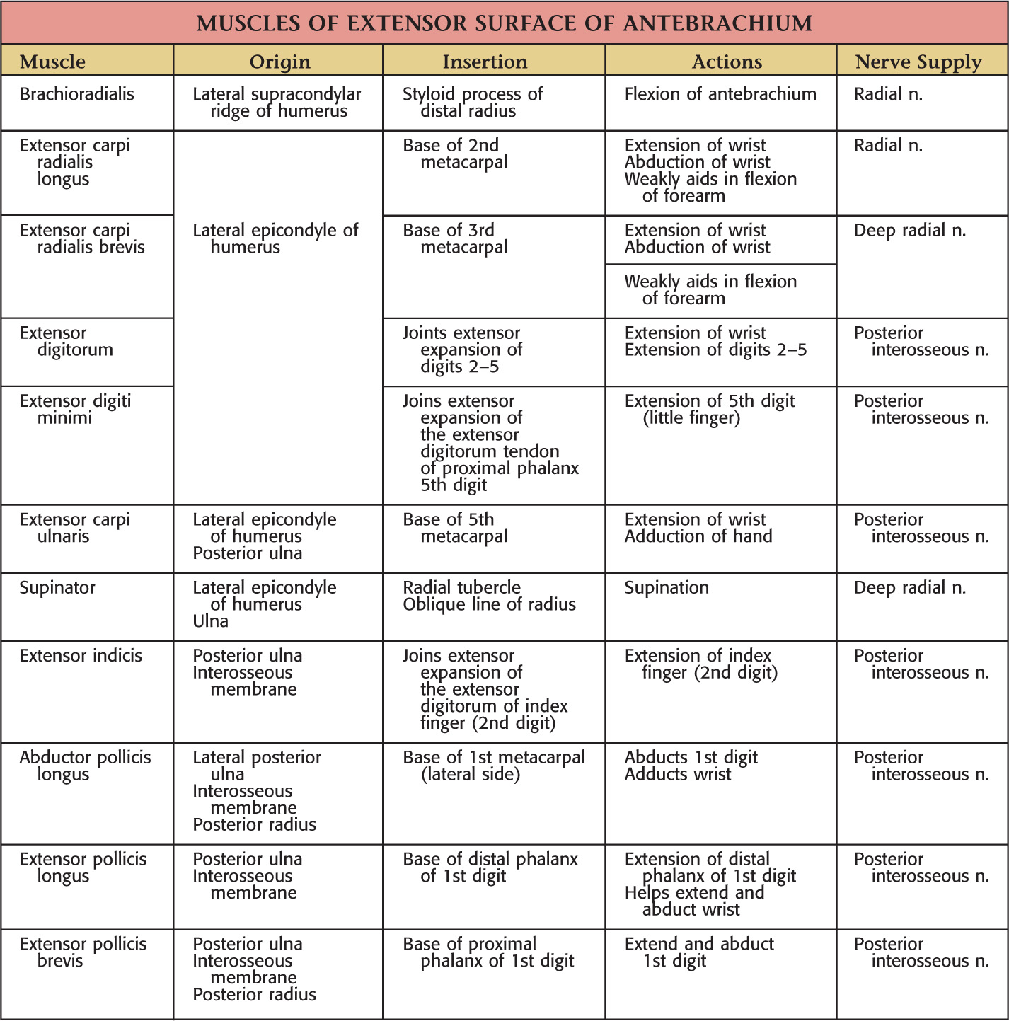
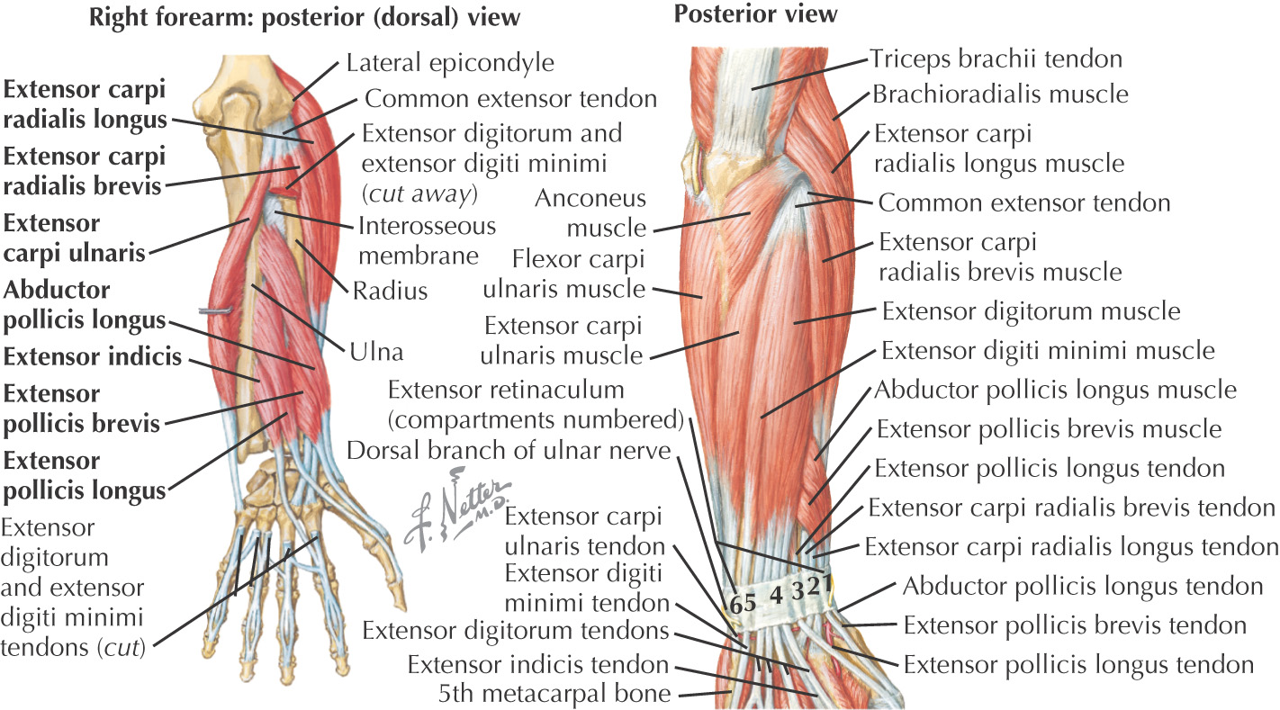
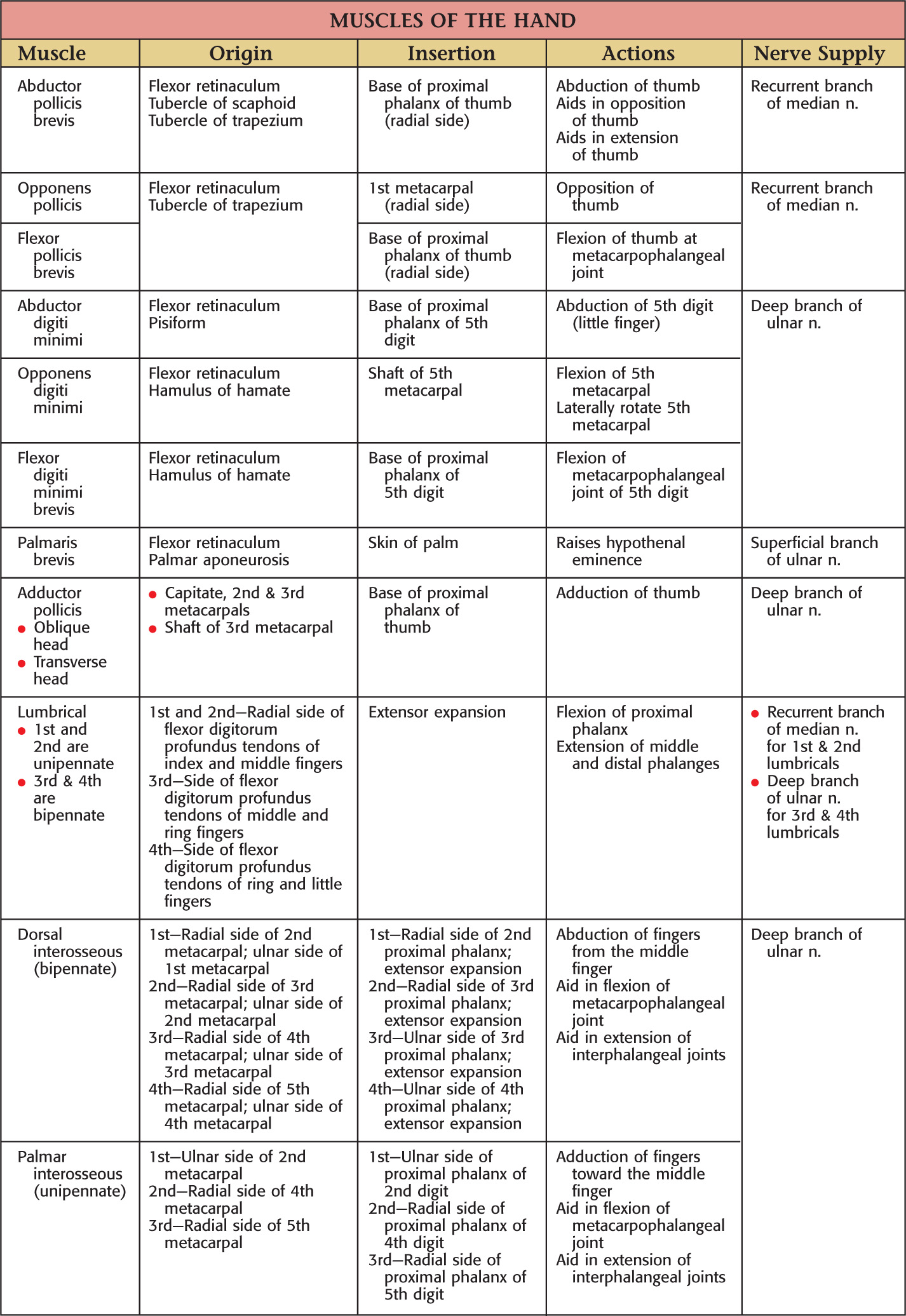
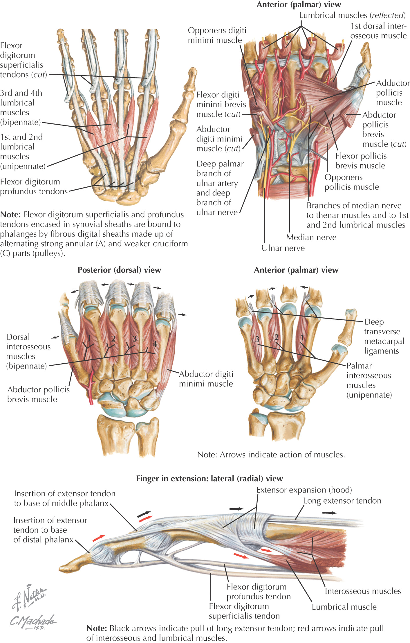
UPPER LIMB
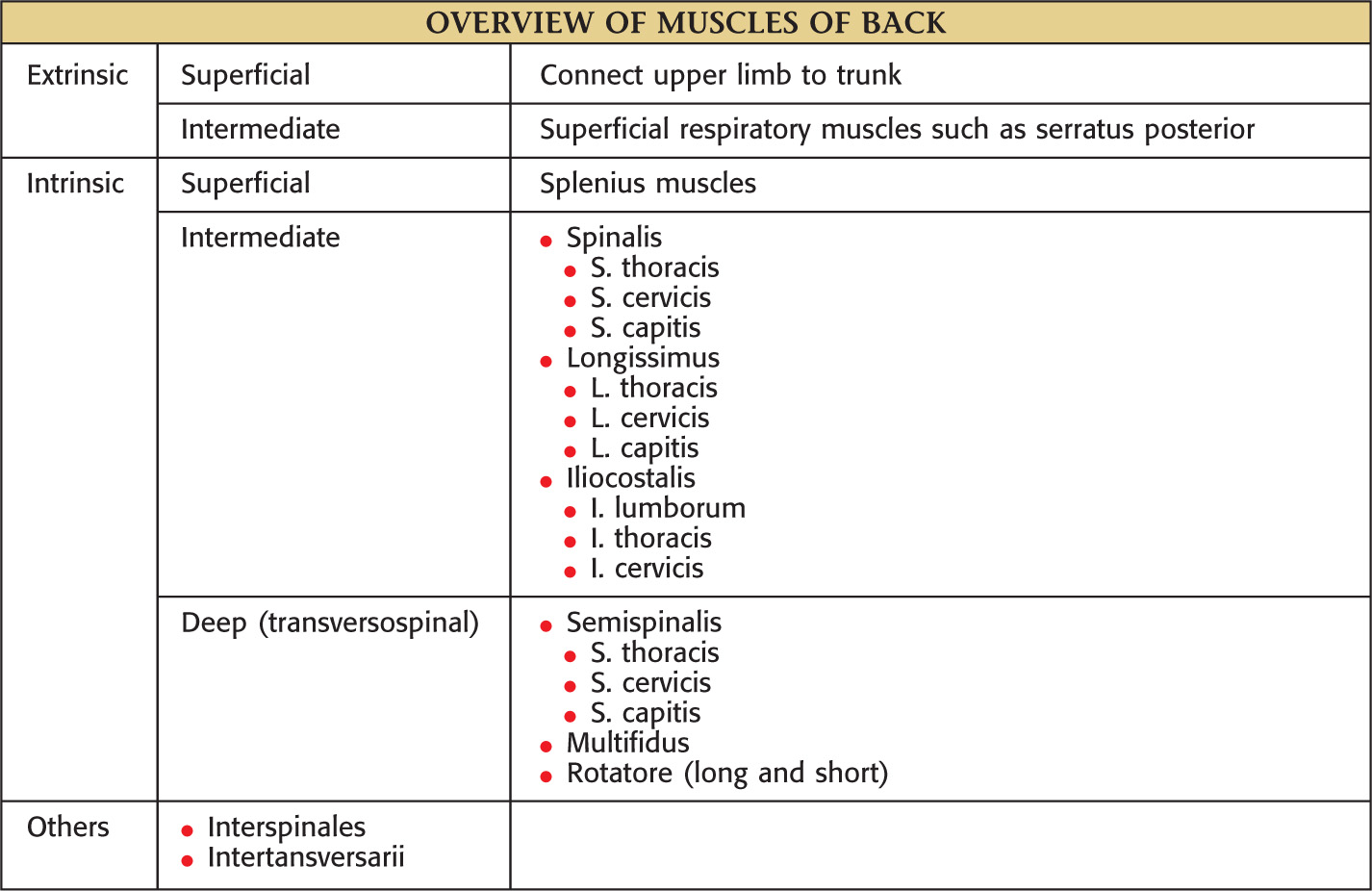
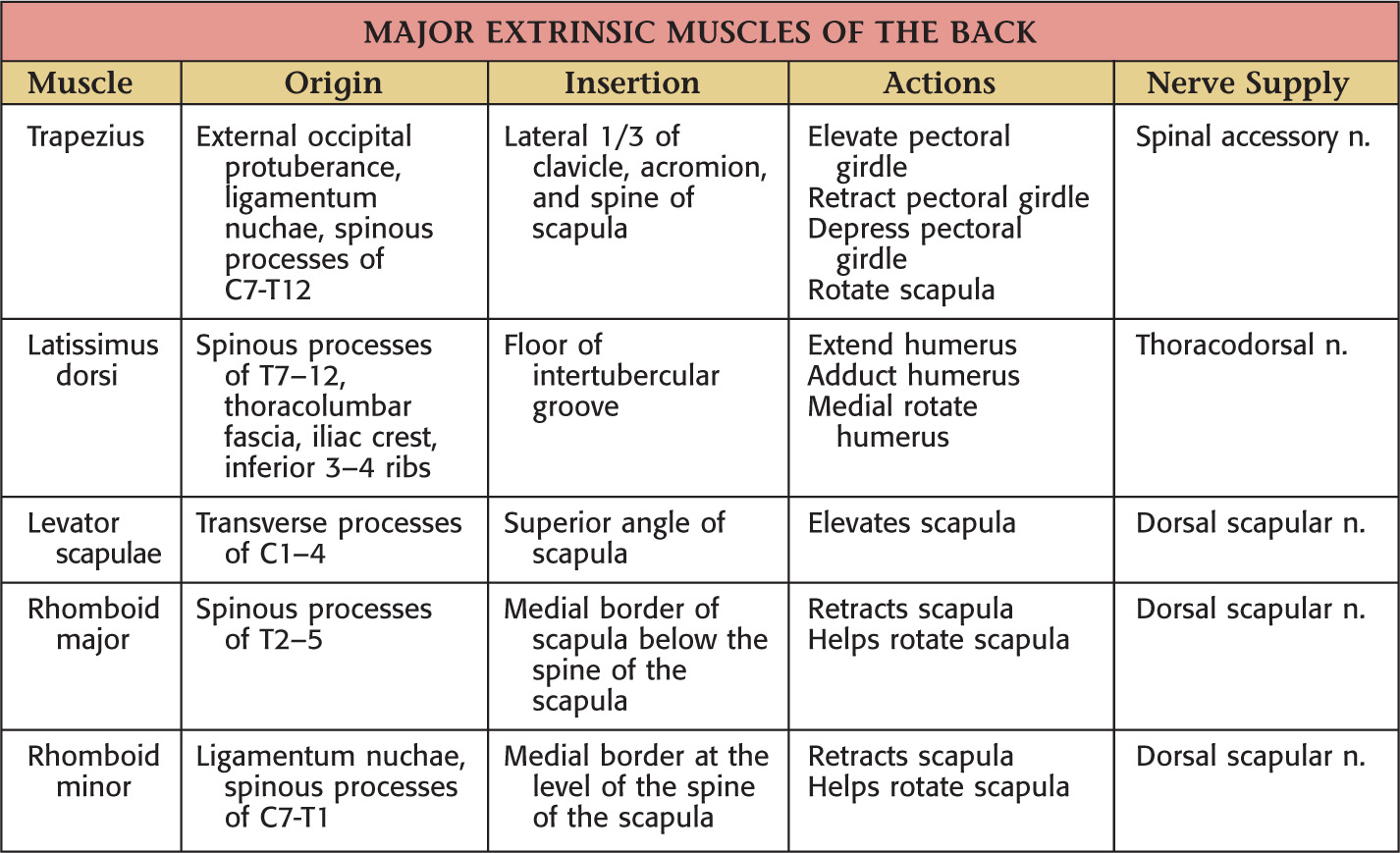
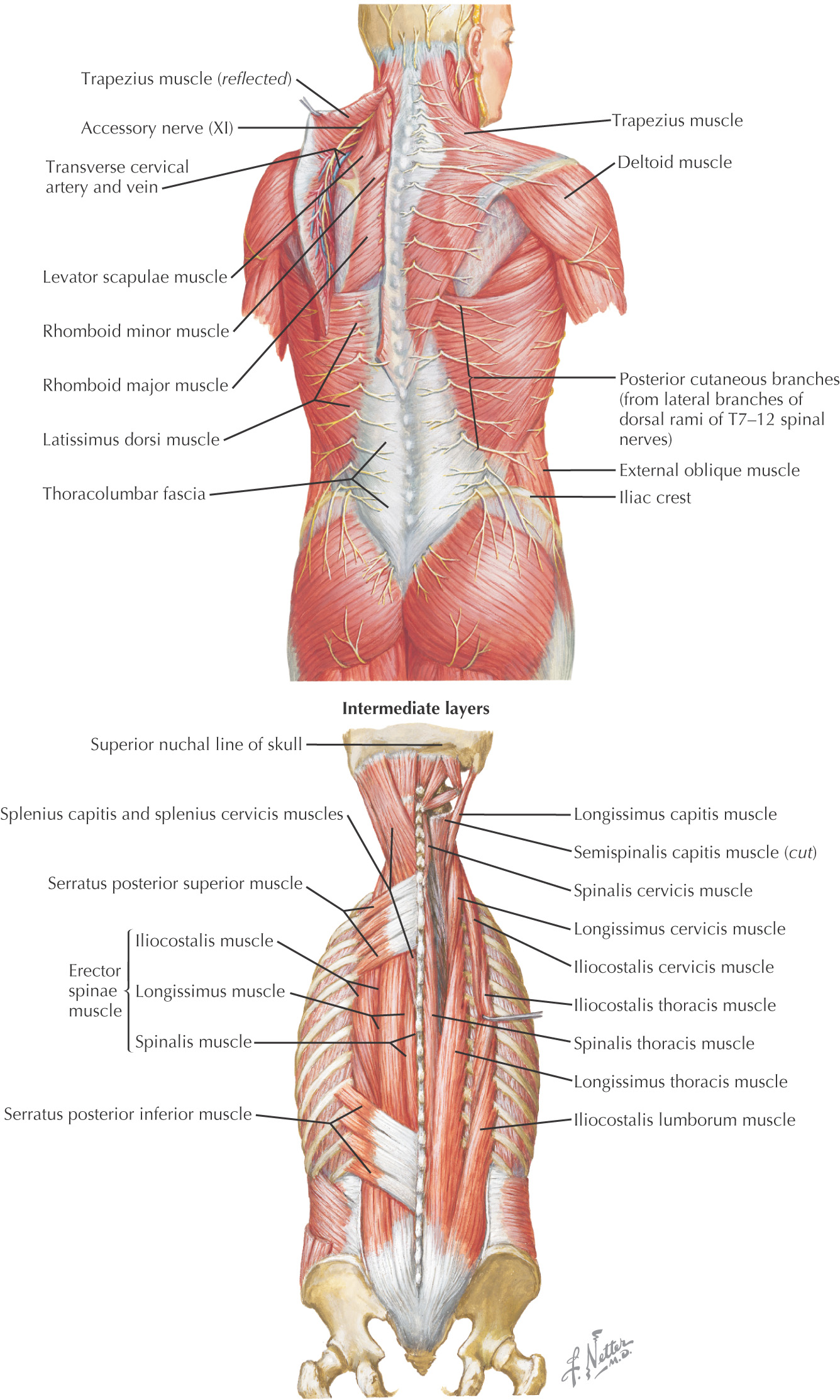
BACK
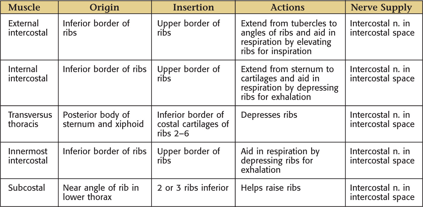
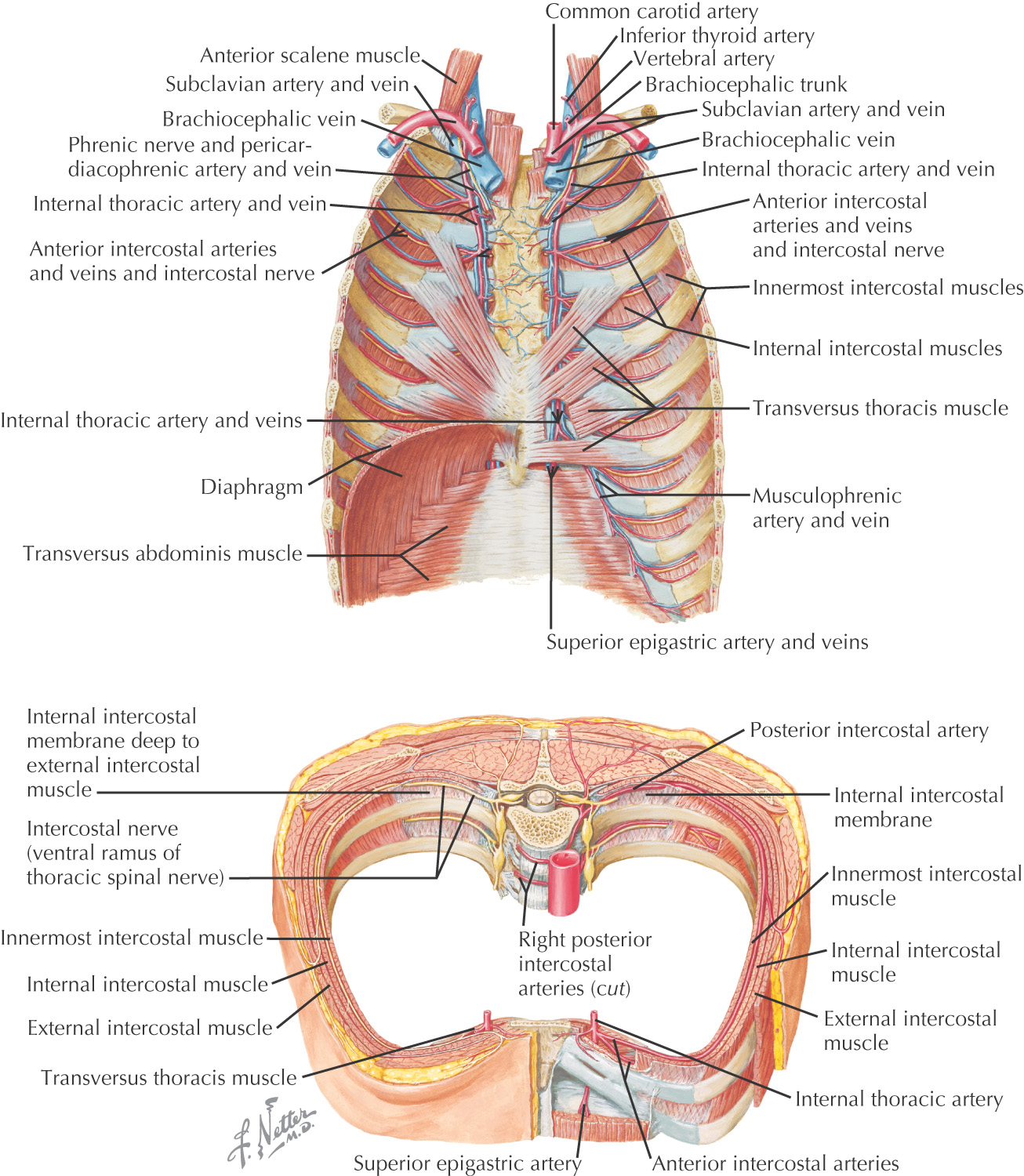
THORAX
ABDOMEN
Contents of the Thorax
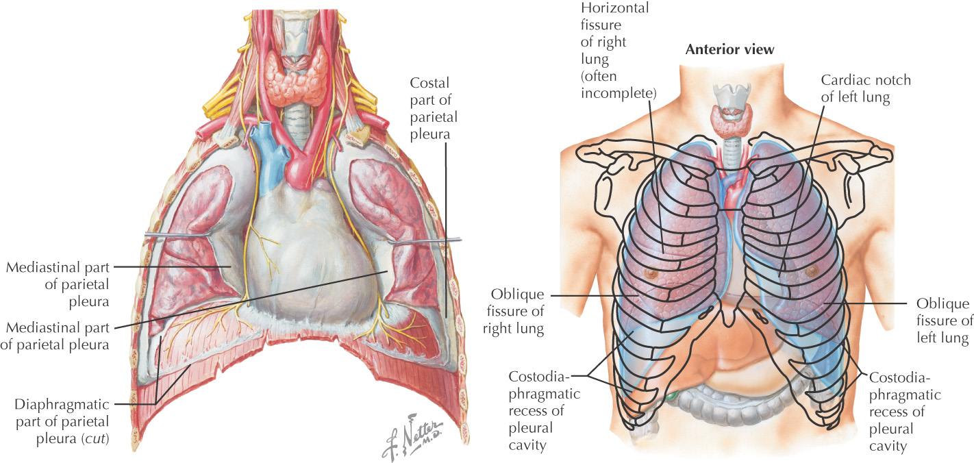
PLEURAL CAVITY
There are 2 pleural cavities
The cavity is composed of a 2-layered pleural sac that secretes a thin layer of serous fluid
• Visceral layer—lines the lung and fissures
• Parietal layer—lines the wall of the cavity
• Costal—lines the cavity along the ribs
• Mediastinal—lines the cavity along the mediastinum
• Diaphragmatic—lines the cavity along the diaphragm
• Cervical (cupula)—lines the cavity forming a dome in the ribs opposite the apex of the lung
Pleural reflections—Abrupt lines where the parietal pleura folds back or changes direction
• Costal (inferior)—where costal pleura is continuous with diaphragmatic pleura
Boundaries
• Anterior midline—6th rib (right) 4th rib (left)
Inferior border of lungs in quiet respiration
• Anterior midline—6th rib (right) 4th rib (left)
Pleural recesses—potential spaces in the pleural cavity where parts of the parietal pleura contact one another during quiet respiration
• Costomediastinal—potential space where costal and mediastinal pleurae come together
• Costodiaphragmatic—potential space where costal and diaphragmatic pleurae come together
Pulmonary ligament—a fold created where the mediastinal pleura at the root of the lung come together and extend inferiorly
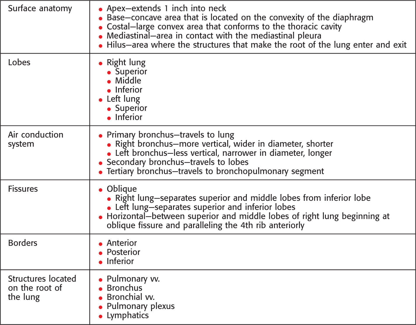
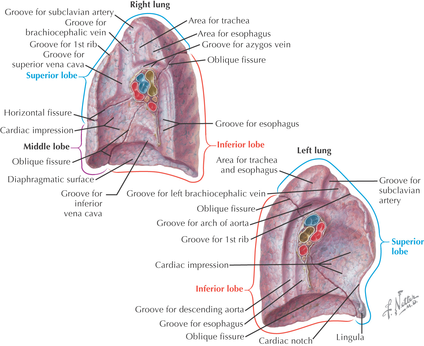
LUNGS
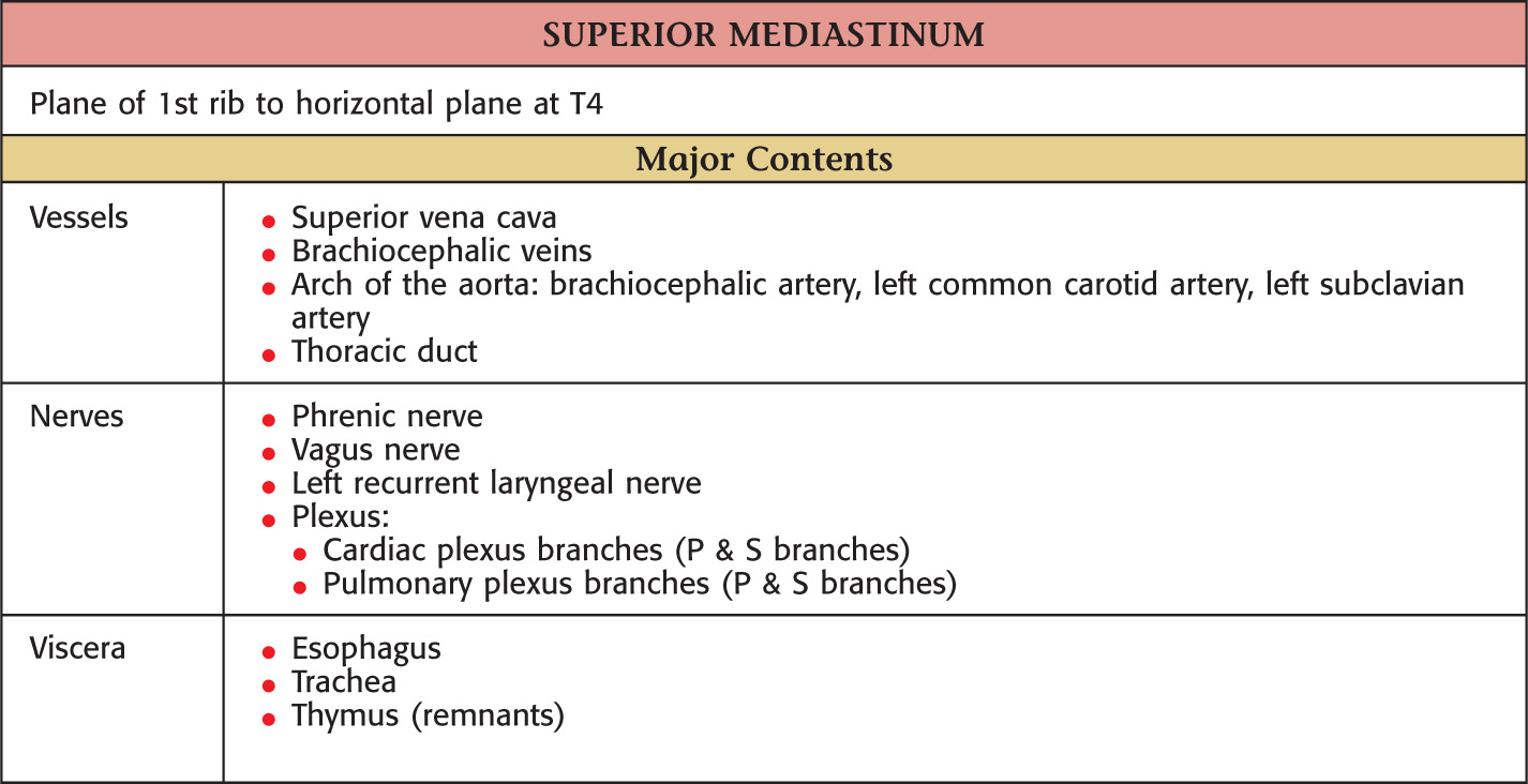
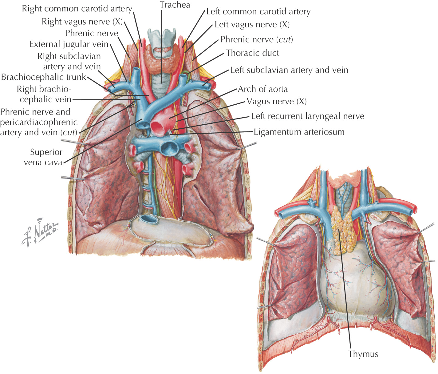
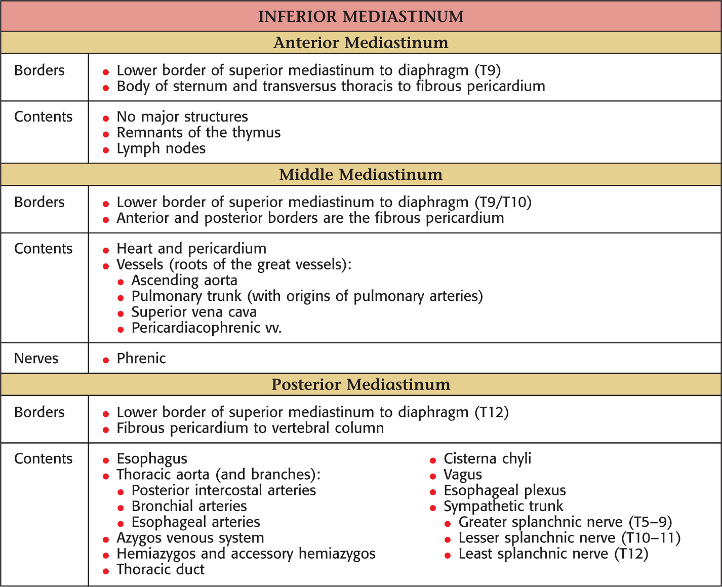
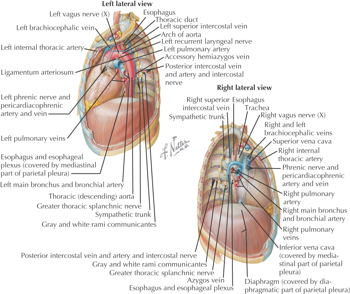
MEDIASTINUM
Region in the middle of the thorax between the two pleural sacs
Subdivided into superior and inferior
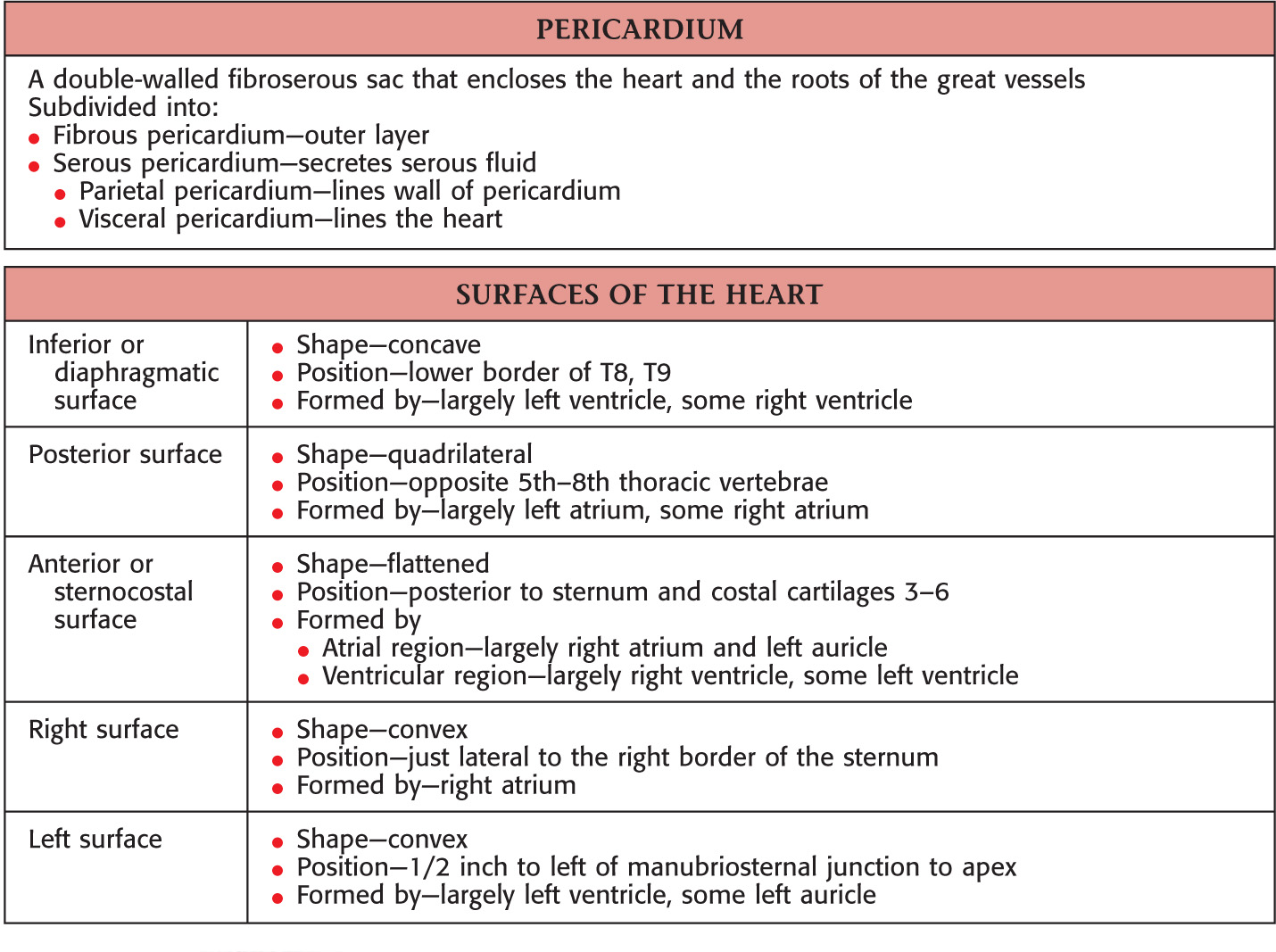
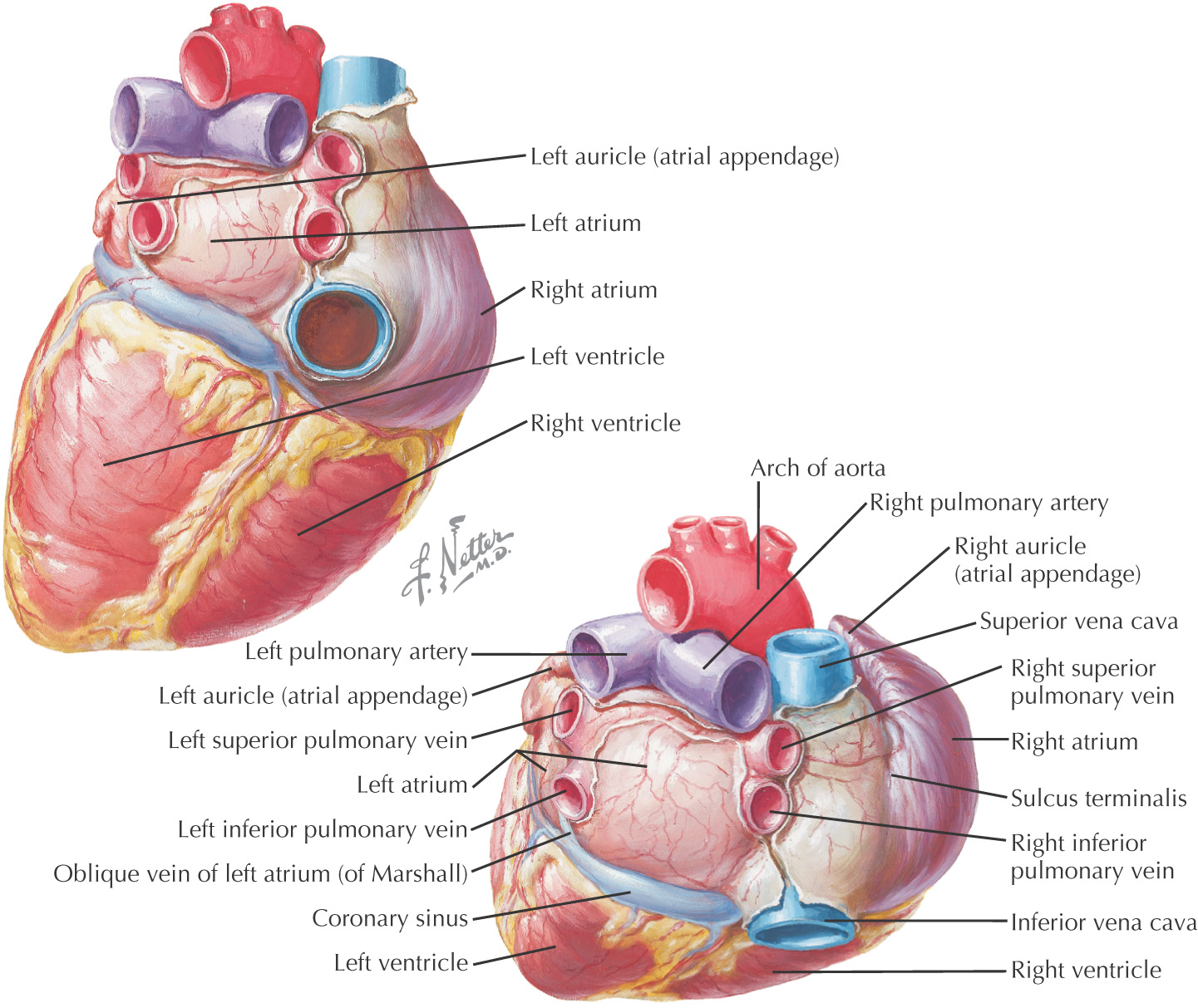
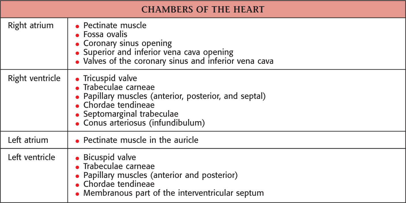
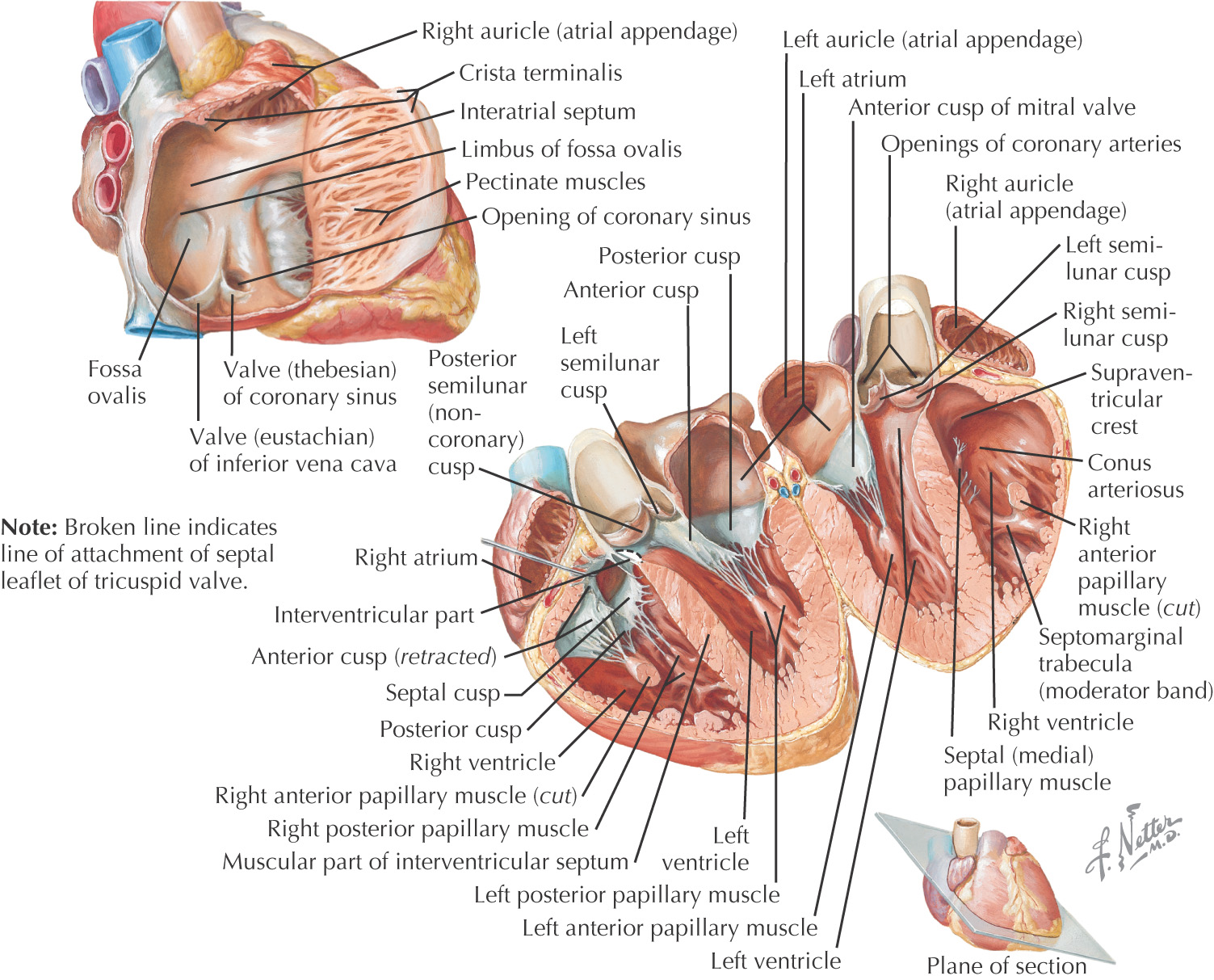
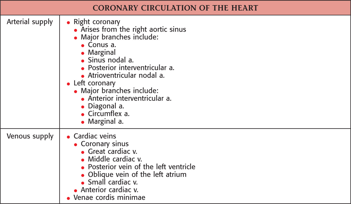
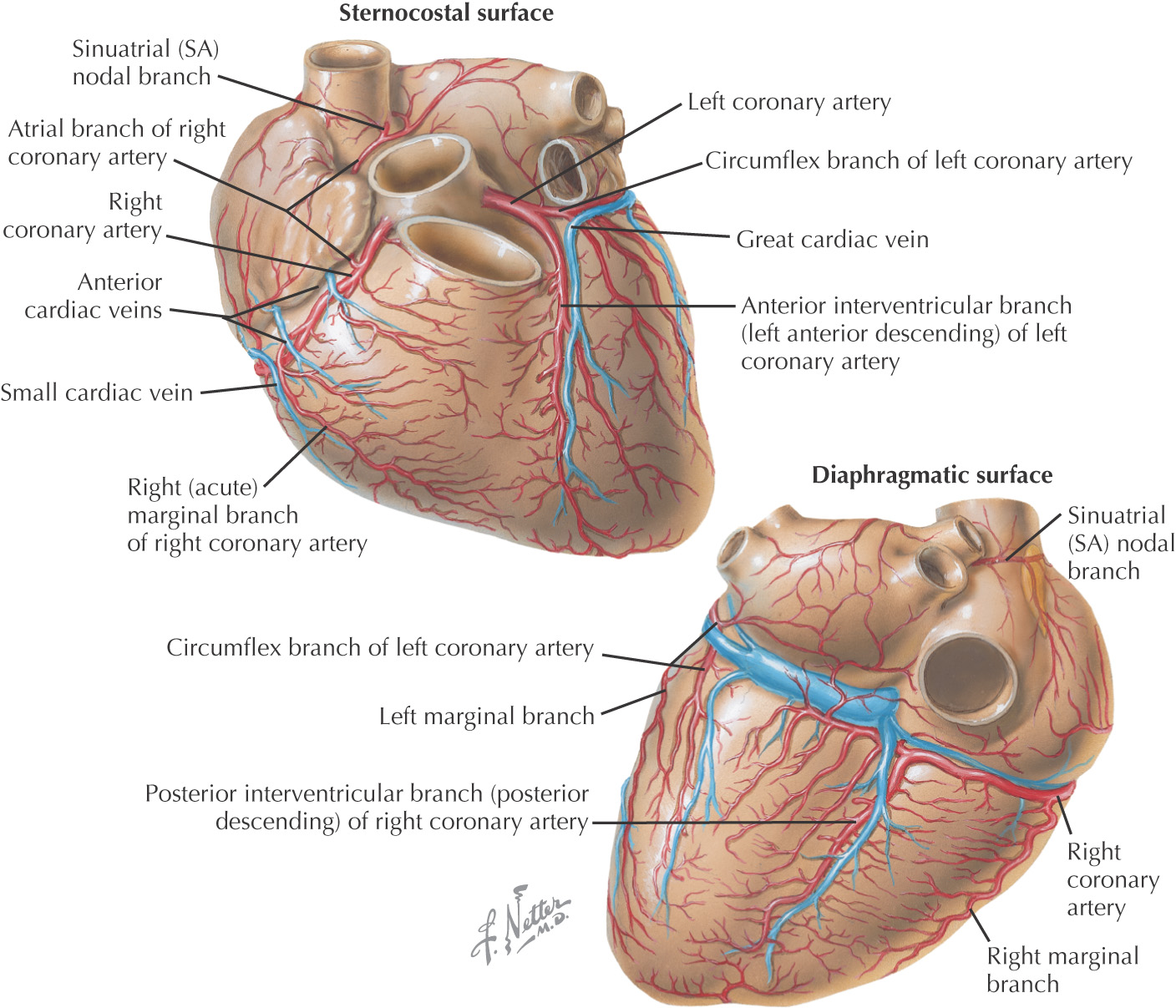
HEART
Contents of the Abdomen
STOMACH
Part of the foregut
There are 4 anatomical parts of the stomach:
• Cardia—where esophagus enters the stomach
• Fundus—created by the superior portion of the greater curvature
• Pylorus—inferior portion that continues to narrow until reaching the pyloric sphincter
The mucosal lining of the stomach are raised elevations known as gastric rugae
There are 2 major curvatures:
• Greater curvature—provides attachment for some remnants of the dorsal mesogastrium:
• Lesser curvature—provides attachment for remnants of the ventral mesogastrium:
• Hepatogastric portion (hepatoduodenal portion does not attach to the stomach)
There are 2 sphincters associated with the stomach:
• Esophageal—not an anatomical sphincter
Stay updated, free dental videos. Join our Telegram channel
VIDEdental - Online dental courses


