Introduction
The purpose of this cross-sectional retrospective study was to evaluate the patterns of pharyngeal airway volume change determined by cervical vertebral maturation (CVM) stage and compare it with that which was characterized by chronological age. Correlations between hyoid bone positions and airway volumes were also examined.
Methods
CVM staging was determined from cone-beam computed tomography scans of 420 white patients aged 9-15 years. Patients were stratified on the basis of sex and skeletal pattern to establish pharyngeal airway volume clusters for each CVM stage. The horizontal and vertical positions of hyoid bones were measured using Hyoidius and Sella.
Results
Males had larger pharyngeal airway volumes compared with females. In males, the largest increases in pharyngeal airway volumes occurred at an earlier CVM stage than females. No statistically significant differences in pharyngeal airway volumes were noted in subjects with skeletal Class I, II, and III malocclusion. The hyoid bone in males was more anteriorly and inferiorly positioned compared with females. The Class III group had a further forward position of the hyoid bone than the Class I and II groups.
Conclusions
The patterns of pharyngeal airway change obtained using CVM staging did not correlate well with traditional maturational models for skeletal growth. It implies that chronologic age could be a relatively reliable indicator for the assessment of pharyngeal airway volumes in adolescents, as outlined in part 1 of the present study. Subjects with anteriorly and superiorly positioned hyoid bones exhibited smaller pharyngeal airway volumes, which highlights the role of soft tissue and its influence on airway patency.
Highlights
- •
Patterns of pharyngeal airway change from CVM staging was compared with chronological age.
- •
The hyoid bone positions also were examined using CBCTs from 420 Caucasian adolescents.
- •
CVM staging showed questionable validity for pharyngeal airway volume assessment.
- •
The hyoid bone moved inferiorly as the maturation of cervical vertebrae progressed.
- •
A notably forward-positioned hyoid bone could imply soft tissue impingement on the airway.
Identification of the skeletal maturity in adolescents has been considered an essential component of orthodontic diagnosis. Because chronological age presented a moderate level of prediction for skeletal maturation status, various maturational indicators have been proposed to determine the proper timing for growth modification therapy in growing patients. Of these, the use of hand-wrist radiographs (HWR), developed by Fisher in the 1980s, is still the gold standard to assess skeletal maturity and growth. , However, although this is a reliable method, it requires additional radiation exposure to the patient, which goes against the principles of as low as reasonably achievable for ionizing radiation. As a result, an alternative to hand-wrist radiographs was developed on the basis of the cervical vertebral maturation (CVM) on cephalograms. Since its conception, multiple authors have attempted to compare the CVM method with HWR to assess the reliability and validity with conflicting results. ,
The present study is the second of 2 articles describing the findings of a cross-sectional retrospective study on pharyngeal airway volumes as determined from cone-beam computed tomography (CBCT). The first presented the results of chronological assessments; the second examined the results obtained from CVM assessments in conjunction with hyoid bone measurements.
The close relationship between the pharynx and hyoid bone justifies orthodontic interest to study their interactions. Hyoid bone is the only bone not articulated to any other bone in the body and is connected to the pharynx, mandible, and cranium through muscles and ligaments. Previous studies suggested a forward and upward displacement of the hyoid bone after mandibular advancement surgery or through the use of functional appliances for mandibular repositioning. , Although there is abundant evidence regarding hyoid bone position after interceptive orthodontic or orthognathic therapies, information about hyoid bone and airway volumes in untreated patients remains scarce.
The purpose of this study was to evaluate the patterns of pharyngeal airway volume change determined by CVM staging and compare it with that which was characterized by chronological age. Another goal of this study was to assess the impact of the location of the hyoid bone on the pharyngeal airway volume of children aged 9-15 years. The null hypothesis is that there are no interactions among skeletal pattern, sex, and CVM stage in the pharyngeal airway volumes. Furthermore, there is no correlation between pharyngeal airway volume and hyoid bone position.
Material and methods
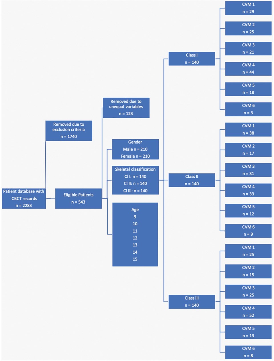
This cross-sectional retrospective study is a follow up to the study of Chan et al with the inclusion of CVM staging and hyoid bone measurements as part of the airway analyses. CBCTs from 420 patients’ initial records at the University of Detroit Mercy Orthodontic Clinic were examined for this project (210 males and 210 females), with ages ranging from 9-15 years. No CBCTs were taken for the purpose of this present study. It was approved by the corresponding Institutional Review Board, number 1819-48. The process of subject redistribution with CVM staging was outlined in Figure 1 . Details of the experimental design, study groups, and methods were published in the earlier article.
The CVM staging protocol developed by Baccetti et al and McNamara Jr et al was adopted for use in this study. The morphology of the bodies of the second (odontoid process, C2), third (C3), and fourth (C4) cervical vertebrae was analyzed visually for CVM determination. CVM stage was determined twice by the same investigator. If there were inconsistent findings between 2 consecutive observations on the CVM stage, a consensus decision was made with another orthodontist.
Each digitized i-CAT image was oriented parallel to the Frankfurt Horizontal plane (FHP) for analysis of the hyoid bone. Hyoidius, the most anterior and superior point of the hyoid bone, was used as a cephalometric landmark because it is the most reproducible. , The position of the hyoid bone was determined in both the sagittal and vertical planes. The horizontal reference plane consisted of a line passing through Sella parallel to the FHP (Sella horizontal line), whereas the vertical reference plane was a line passing through Sella perpendicular to FHP (Sella vertical line). Hyoid bone vertical (HBV) and hyoid bone horizontal (HBH) measurements were taken by drawing a line from the Hyoidius of the hyoid bone perpendicular to the reference lines ( Fig 2 ).

Statistical analysis
Fifty cephalograms were evaluated by 1 investigator (L.V) and categorized into 1 of the 6 CVM stages. After a wash-out period of 2 weeks, the same investigator reevaluated all images. The reliability of CVM determination was confirmed using the weighted kappa intraclass correlation test. Pearson correlation analysis was used to detect the correlation between CVM stage and airway dimensions. A P <0.05 was set to be statistically significant. All analyses were performed using SAS software (version 9.4; SAS Institute, Cary, NC). An analysis of variance (ANOVA) was used to compare airway volumes within CVM stages and between other parameters such as sex and skeletal pattern followed by the post-hoc test to identify significant differences among the 6 CVM groups. Hyoid bone measurements were subjected to the same statistical analyses. Pearson correlation was used to evaluate the correlation between hyoid bone measurements and airway volumes.
Results
According to the weighted kappa intraclass correlation and Pearson correlation tests, the concordance index was greater than 0.99 for hyoid bone measurements ( Table I ). For CVM staging, the linear weighted kappa statistic was 0.897, indicating high intraexaminer concordance ( Table I ). Descriptive statistics for the CVM stage, skeletal pattern, sex, and pharyngeal airway volume were outlined in Table II .
| Variable | Kappa | Pearson correlation |
|---|---|---|
| Cervical vertebral maturation stage | 0.897 | × |
| HBV | × | 0.998 |
| HBH | × | 0.997 |
| SNA | SNB | ANB | NPA (mm 3 ) | OPA (mm 3 ) | TPA (mm 3 ) | |||||||||
|---|---|---|---|---|---|---|---|---|---|---|---|---|---|---|
| CVM | Skeletal pattern | Sex | Mean | SD | Mean | SD | Mean | SD | Mean | SD | Mean | SD | Mean | SD |
| 1 | I | Female | 80.7 | 3.1 | 77.0 | 2.8 | 3.7 | 0.8 | 3351.1 | 1879.5 | 5685.0 | 1454.0 | 9036.1 | 2844.8 |
| Male | 80.5 | 3.7 | 77.5 | 3.8 | 3.0 | 1.2 | 4091.6 | 1812.8 | 7834.5 | 4282.1 | 11926.1 | 5445.2 | ||
| II | Female | 79.8 | 2.1 | 73.6 | 2.2 | 6.2 | 1.3 | 4471.7 | 2729.0 | 6647.5 | 2200.2 | 11119.1 | 4468.9 | |
| Male | 82.7 | 2.9 | 76.2 | 2.5 | 6.5 | 1.2 | 4808.6 | 2008.8 | 7461.4 | 2883.6 | 12270.0 | 4238.3 | ||
| III | Female | 78.3 | 3.1 | 78.1 | 3.6 | 0.3 | 0.9 | 2698.5 | 1045.3 | 6234.1 | 3424.6 | 8932.6 | 3989.6 | |
| Male | 77.9 | 4.4 | 78.6 | 4.5 | −0.7 | 1.2 | 3590.7 | 1690.3 | 8816.4 | 3471.9 | 12407.1 | 4169.2 | ||
| 2 | I | Female | 81.1 | 3.5 | 78.0 | 3.3 | 3.1 | 1.1 | 4425.8 | 1437.4 | 8967.6 | 3358.0 | 13393.3 | 4241.6 |
| Male | 81.3 | 3.9 | 78.2 | 3.7 | 3.1 | 1.2 | 4960.5 | 1752.0 | 9803.0 | 2974.1 | 14763.4 | 4385.4 | ||
| II | Female | 82.4 | 2.1 | 75.9 | 2.7 | 6.5 | 1.4 | 3141.4 | 1047.5 | 6840.2 | 1766.4 | 9981.6 | 1912.8 | |
| Male | 81.9 | 3.3 | 75.2 | 3.6 | 6.7 | 1.5 | 4374.0 | 1385.7 | 8782.2 | 5782.5 | 13156.3 | 6376.0 | ||
| III | Female | 78.5 | 3.3 | 79.7 | 3.3 | −1.2 | 1.2 | 3837.1 | 1249.2 | 8083.3 | 3408.8 | 11920.4 | 4134.0 | |
| Male | 79.4 | 3.2 | 80.4 | 3.4 | −1.0 | 0.8 | 5081.9 | 1656.1 | 9961.5 | 4086.4 | 15043.4 | 5623.2 | ||
| 3 | I | Female | 80.9 | 2.0 | 77.5 | 2.1 | 3.4 | 1.3 | 4559.2 | 1496.8 | 8780.6 | 2641.7 | 13339.9 | 3812.8 |
| Male | 81.0 | 3.4 | 77.4 | 3.8 | 3.6 | 1.3 | 4525.7 | 1509.1 | 9680.2 | 3384.9 | 14205.9 | 4359.0 | ||
| II | Female | 83.2 | 3.6 | 76.9 | 3.6 | 6.2 | 1.4 | 4355.6 | 1965.7 | 9076.0 | 3694.7 | 13431.6 | 4992.0 | |
| Male | 83.1 | 3.2 | 77.2 | 4.8 | 6.4 | 1.1 | 4987.1 | 2154.6 | 9753.5 | 3604.7 | 14740.6 | 5014.5 | ||
| III | Female | 78.5 | 3.5 | 78.7 | 3.6 | −0.2 | 1.0 | 4759.1 | 1461.0 | 11587.7 | 3641.1 | 16346.7 | 4445.6 | |
| Male | 79.7 | 2.0 | 80.1 | 2.4 | −0.4 | 1.1 | 5240.8 | 1932.7 | 8996.1 | 4042.3 | 15136.9 | 5386.7 | ||
| 4 | I | Female | 81.1 | 3.4 | 78.0 | 3.1 | 3.1 | 1.1 | 4138.4 | 1506.9 | 8808.6 | 3579.7 | 12947.0 | 3971.4 |
| Male | 82.5 | 3.9 | 79.4 | 4.2 | 3.1 | 0.9 | 4772.9 | 1854.8 | 13364.1 | 6029.0 | 18137.0 | 7197.6 | ||
| II | Female | 82.6 | 3.0 | 76.1 | 3.1 | 6.5 | 1.6 | 3956.9 | 1397.3 | 9480.8 | 4131.4 | 13427.7 | 4775.1 | |
| Male | 83.3 | 3.8 | 77.1 | 3.5 | 6.2 | 1.0 | 5878.9 | 1685.6 | 12447.4 | 5442.4 | 18326.3 | 6424.0 | ||
| III | Female | 79.4 | 3.2 | 80.6 | 3.3 | −1.3 | 1.8 | 4489.0 | 2612.5 | 11120.0 | 5861.5 | 15609.0 | 7250.6 | |
| Male | 80.3 | 4.4 | 81.0 | 4.8 | −0.7 | 1.6 | 4964.6 | 2162.3 | 11875.6 | 6176.3 | 16840.1 | 7897.7 | ||
| 5 | I | Female | 81.0 | 3.0 | 78.5 | 2.9 | 2.6 | 1.0 | 5338.7 | 1430.0 | 11827.5 | 4269.6 | 17166.2 | 4994.0 |
| Male | 81.9 | 2.1 | 78.8 | 2.5 | 3.1 | 1.0 | 5937.8 | 2562.3 | 12724.4 | 1930.1 | 18662.2 | 3131.2 | ||
| II | Female | 83.6 | 3.9 | 76.7 | 4.7 | 6.9 | 1.4 | 4864.1 | 2516.3 | 12544.5 | 5428.8 | 17408.6 | 6385.2 | |
| Male | 84.5 | 5.8 | 77.1 | 4.6 | 7.4 | 2.4 | 6278.4 | 984.3 | 12488.0 | 4771.9 | 18766.4 | 5678.9 | ||
| III | Female | 80.2 | 4.9 | 81.8 | 4.2 | −1.6 | 2.2 | 4777.1 | 2333.7 | 12997.6 | 8241.3 | 17774.7 | 10317.1 | |
| Male | 80.2 | 3.5 | 83.7 | 1.1 | −3.6 | 4.5 | 5843.1 | 702.8 | 21114.7 | 9755.7 | 26957.8 | 10458.5 | ||
| 6 | I | Female | 79.2 | 1.7 | 76.9 | 0.9 | 2.3 | 0.8 | 5651.0 | 2394.5 | 13279.5 | 6796.5 | 18930.6 | 9158.7 |
| Male | – | – | – | – | – | – | – | – | – | – | – | – | ||
| II | Female | 83.6 | 3.9 | 77.0 | 4.4 | 6.6 | 1.1 | 5642.8 | 1303.2 | 10327.4 | 2462.7 | 15970.2 | 3190.6 | |
| Male | 86.5 | – | 80.0 | – | 6.5 | – | 9822.9 | – | 19249.8 | – | 29072.7 | – | ||
| III | Female | 81.6 | 3.7 | 83.2 | 3.0 | −1.6 | 3.1 | 5372.9 | 2108.3 | 10095.5 | 1763.6 | 15468.3 | 2356.7 | |
| Male | 81.1 | – | 83.8 | – | −2.8 | – | 3915.7 | – | 14441.1 | – | 18356.8 | – | ||
Stay updated, free dental videos. Join our Telegram channel
VIDEdental - Online dental courses


