Chapter 6
Oral embryology and histology
DEFINITIONS
Oral embryology is the study of the development of the oral cavity, and the structures within it, during the formation and development of the embryo in the first 8 weeks of pregnancy.
After this point, the unborn child is referred to as a foetus.
The correct biological term for this time period before birth is prenatal.
Histology is the specialised biological area of study concerned with the microscopic structure and function of tissues.
This chapter will cover the embryological development of all oral structures, as well as the basic histology of the embryonic tissues.
The histology of the following dentally relevant structures warrant more detailed notes, and are discussed elsewhere:
- Tooth histology – covered in Chapter 8
- Periodontal histology – covered in Chapter 9
- Salivary gland histology – covered in Chapter 10
OVERVIEW OF GENERAL EMBRYO DEVELOPMENT
To understand the embryological development of the oral tissues, the dental care professional (DCP) needs to have a basic knowledge of the process of embryo formation, and the significance of the origins of various body tissues and organs to help understand how congenital defects, pathology and disease processes occur.
The 8-week time span of the embryo is summarised in the following text.
Following fertilisation of the female egg by male sperm, the resultant single cell, or zygote, travels to the womb (uterus) while undergoing repeated cell divisions that double the number of cells present; from one to two, then to four, then eight, and so on.
At about 6 days after fertilisation, the cluster of cells so produced then forms into two distinct groups, with the outer layer of cells forming a wall around the inner group, and they develop as follows:
- Outer wall – attaches the cell group into the lining of the uterus, and forms the placenta
- Inner cluster – expands to form the embryo
The inner cells then organise themselves into a flat disc, eventually containing three distinct layers of cells (and one sub-layer) from which all the tissues and organs will develop:
- Ectoderm layer – outer layer of cells, which form the outer skin layer, the sensory cells of the eyes, ears and nose, some of the nervous system and external skin glands
- Neuroectoderm cells – specialised cells from the ectoderm layer, and which form connective tissue, cartilage, some bone and some dental tissues
- Mesoderm layer – middle layer of cells, which form the deep skin layer, muscle, bone marrow and blood cells, lymphatic system, reproductive organs and excretory organs including the salivary glands
- Endoderm layer – inner layer of cells, which form the linings of the respiratory and digestive systems, liver and pancreas
The space between this disc and the outer wall of cells develops as the amniotic sac.
The development of the embryo from weeks 3 to 8 is then rapid and astounding, and is summarised in the following text.
Third week – see Figure 6.1
Figure 6.1 Third-week embryo.
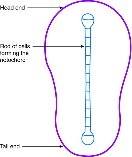
- Disc of cells becomes pear shaped and develops a head end and a tail end
- Rod of cells develops along the back of the embryo to form the notochord, which develops into the spine
- Embryo now has two symmetrical halves – left and right
- Notochord rolls to form a tubular structure that later develops into the brain and spinal cord
Fourth week – see Figure 6.2
Figure 6.2 Fourth-week embryo.
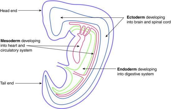
- Cells along the back of the embryo grow more rapidly than the front, forming a curved shape
- Endoderm tissue buds begin developing, and will later form the respiratory and digestive organs
- Brain begins to develop, as do eye stalks and ear pits
- Ectoderm tissue buds develop, and will later form the limbs
- Ectoderm tissue folds develop as branchial arches, which will become the jaws and other neck structures (in lower life forms such as fish, these structures develop as gills)
- Heart begins developing on the front of the embryo, beneath the head, and is pushed down into the chest as the branchial arches develop
Fifth week – see Figure 6.3
- Nose begins developing as nasal pits
- Jaws and ears form
- Hands and feet begin developing
- All internal organs are developing
- Ectoderm tissue folds grow to join at the front and form the chest and abdominal cavity walls
Figure 6.3 Fifth-week embryo.
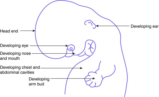
Sixth week – see Figure 6.4
- Eyes and ears are formed
- Mouth and nose begin to develop
Figure 6.4 Sixth-week embryo.
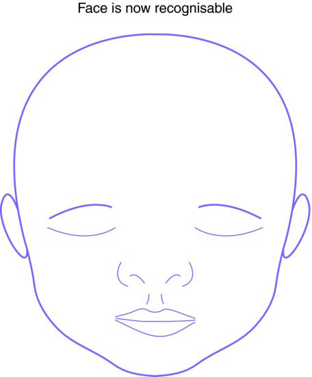
Eighth week – see Figure 6.5
- Face is recognisable
- Embryo curvature relaxes
- Limbs become jointed
- Most internal organs are formed
Figure 6.5 Eighth-week embryo.
ORAL EMBRYOLOGY – OVERVIEW
As summarised previously, the majority of oral embryological development occurs between weeks 3 and 8 after conception, and the oral and facial structures are fully formed by week 12.
As the roof of the mouth, the palate, forms the floor of the nasal cavity, the development of these two cavities is inextricably linked and will be discussed together.
Third week
- Shallow indentation of the ectoderm at the head end of the embryo develops – this is the primitive mouth, or stomodeum
- This is separated from the rudimentary pharynx by a temporary structure called the oropharyngeal membrane
Fourth week onwards
Table 6.1 Branchial arches and associated derivative structures.
- Ectoderm tissue, with considerable neuroectoderm cells present, folds and develops into the six branchial arches, which form various oral and neck structures (see Table 6.1)
- Oropharyngeal membrane disintegrates so that the stomodeum becomes deeper and gradually forms the oral cavity
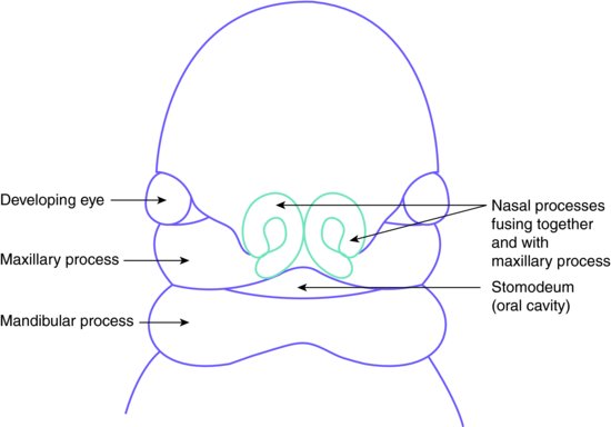
- Two buds of tissue containing cells of all three embryonic layers begin to swell beneath the stomodeum and form the mandibular processes
- Once fused together at the front of the embryo, they form the mandible and its associated structures
- At the same time, two other tissue buds develop from the mandibular processes, and swell above them to form the back section of the maxilla and its associated structures, up to the future position of the canine teeth
- Above the stomodeum, yet another tissue bud develops and forms the frontonasal process
- This goes on to develop the upper face, the nasal structures and the front section of the palate at the future position of the incisor teeth (see Figure 6.6)
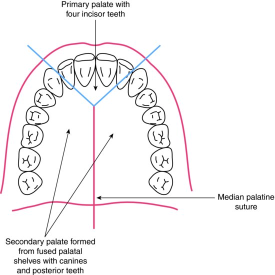
- The front section of the palate is called the primary palate, while the back section is the secondary palate
- The primary palate develops in one piece, while the secondary palate develops as two shelves that join at the midline from week 6 (see Figure 6.7)
Figure 6.6 Early facial development.
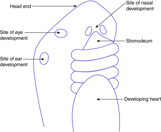
Figure 6.7 The palate.
An understanding of the embryological development of the face and oral cavity helps to explain the cranial nerve supply of the head and neck region, which can otherwise seem a disconcerting area of study to some DCPs.
Embryological development of the tongue
By the fourth week of embryological development, the primitive pharynx has been formed by the first four branchial arches. It is present at this stage as the top end of the hollow tube that will go on to develop into the respiratory and digestive systems. The seal of the oropharyngeal membrane has disintegrated, and the stomodeum is deepening to form the oral cavity.
At this point, tissue buds develop on the floor of the primitive pharynx; those from the first branchial arches develop into the body of the tongue, while those from the second, third and fourth branchial arches eventually develop into the base of the tongue:
- A central tissue bud called the tuberculum impar develops from the first branchial arch
- Two side buds develop next to it as lateral lingual swellings
- These two swellings grow together and encompass the first tissue bud, and the resultant organ becomes the body of the tongue, forming the anterior two-thirds of its whole structure
- As the cells along the sides of the lingual swellings disintegrate, the body of the tongue becomes free from the floor of the mouth except for a central tag of tissue beneath, which is the lingual frenum
- From the third and fourth branchial arches, another pair of swellings develop, called the copula, and these form the base of the tongue
- As the copula grow forwards and encompass tissue from the second branchial arch, they merge with the anterior swellings at around week 8
- The point at which they join is visible as a V-shaped groo/>
Stay updated, free dental videos. Join our Telegram channel
VIDEdental - Online dental courses


