Class II/1, crowding, defective 16, 26, 36, and 46
Extraction 16, 26, 36 and 46, cervical headgear
Female: 10 years, 9 months to 25 years, 7 months
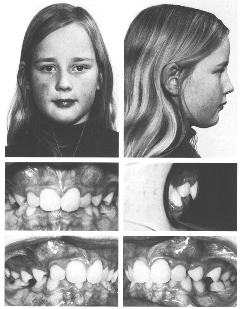
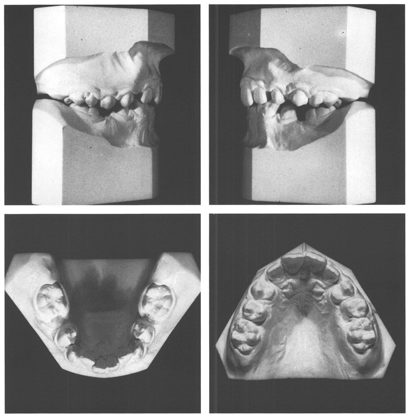
A girl aged 10 years, 0 months displayed a class II/1 anomaly, otherwise having normal function. Significant crowding had developed after premature loss of deciduous molars. The incisors, particularly the maxillary ones, were steeply inclined. There was hardly any room for the maxillary canines. In the mandible, the crowding was concentrated mostly in region of the impacted second premolars. In habitual occlusion the incisors were in contact, but with an overjet of 3 mm and an overbite of 6 mm. After taking into account the drifting of the posterior teeth, a disto-occlusion of ½ PW between the molars was estimated. The problem was complicated by a high caries activity with the consequences of decalcifications, cavities in the first permanent molars, and probably the early loss of the deciduous teeth (Figs 34.1 and 34.2).


Figs 34-1 and 34-2 A girl aged 10 y, 9 mo with a class II/1 anomaly and marked crowding in both dental arches, which is attributable mainly to the early loss of the deciduous molars. The mandibular incisors and the maxillary ones to a greater degree are tipped back. There is an ALD of –9 mm in the mandible and the crowding is localized primarily in the second premolar region. In the maxilla, the ALD is –10 mm; the first permanent molars have migrated mesially and rotated and there is almost no space for the canines which are located buccally high in the vestibule. The dentition is poorly cared for. Plaque is found in many situations and the gingiva are congested. The first permanent molars have deep caries and decalcification buccally and lingually. Even the maxillary incisors have been attacked, although to a lesser extent.
It was decided to extract all four first permanent molars because due to their poor quality they were most unlikely to be able to be conserved for long. After the second permanent molars had come into occlusion, a cervical headgear would be fitted to the maxillary molars to improve the intermaxillary relationship and to secure more space for the maxillary canines. Depending on the oral hygiene and cooperation of the patient, other appliances might be used in due course.
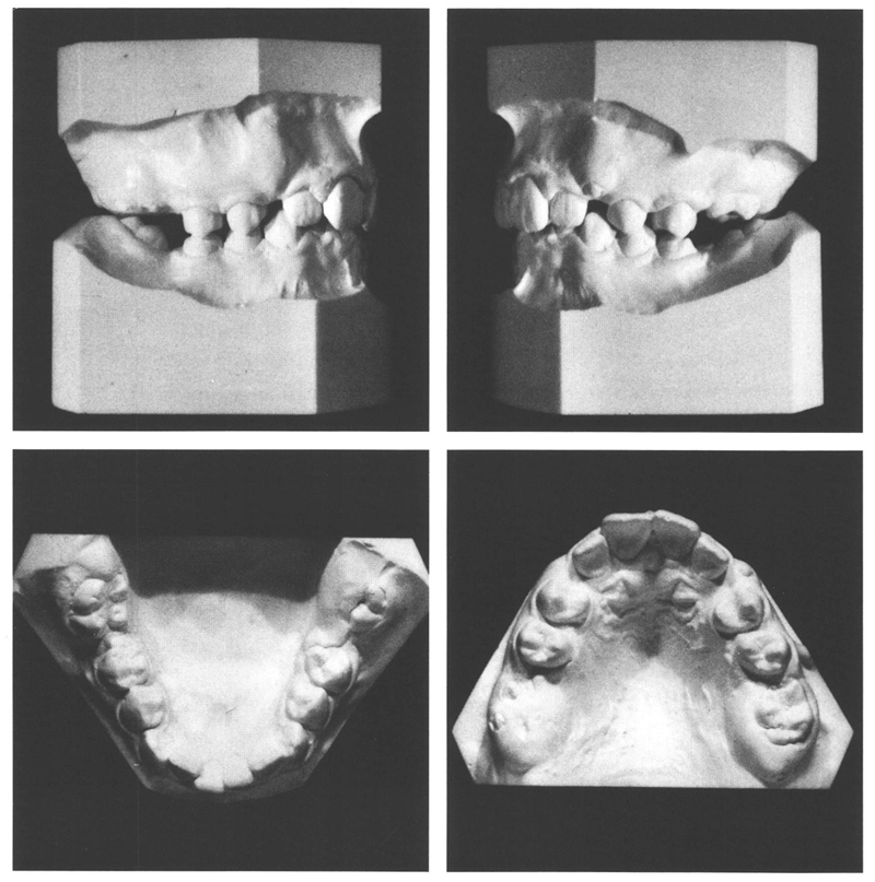
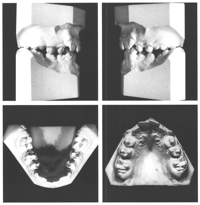
At the age of 10 years, 10 months, the first permanent molars on the right side were removed, and four weeks later, those on the left also were extracted. It is possible to see on the models provided six months later when aged 11 years 5 months, that the mandibular second premolars had emerged with all the second permanent molars (Fig. 34.3). Six months later, the second molars were in occlusion and the space between the second premolars and the second molars in both jaws had diminished, although those teeth were tipped and rotated. The space in the maxilla for the canines had scarcely increased as a result of the extractions and the canines had emerged ectostomatically (Fig 34.4). At this stage, the maxillary second molars were banded and a cervical headgear was fitted. It was the intention to improve the molar occlusion by this means, and meanwhile to correct the rotations of the maxillary second molars and increase the space available for the canines.

Fig 34-3 Six months after extraction of the first permanent molars, the second premolars in the mandible and all the second permanent molars have either fully or partly emerged. The first premolars and the canines in the mandible have drifted a little distally, enabling the lateral incisors to move to labial. In the maxilla, the extraction of the first permanent molars has not contributed much to increasing the room available for the canines. The lateral photographs were not taken perpendicular to the median plane but are perpendicular to the molar/premolar arch segments.

Fig 34-4 A year after the extraction of the first permanent molars, both the mandibular left second molar and that on the right in the maxilla have fully emerged. The space available for the maxillary canines has increased only slightly; the canines have emerged ectostomatically. The mesio-palatally rotated second molars are located close to the second premolars.
After 2½ years, during which the wearing of the headgear was not satisfactory, treatment was conclu/>
Stay updated, free dental videos. Join our Telegram channel

VIDEdental - Online dental courses


