Chapter 2
Cardiovascular system
The cardiovascular system is composed of the following:
- The heart
- Blood vessels carrying blood away from the heart – arteries and arterioles
- Blood vessels carrying blood to the heart – veins and venules
- Microscopic blood vessels connecting these two systems together – capillaries
- Blood – consisting of various cells suspended in a fluid – plasma
The whole system is sealed and enclosed, and runs in a similar fashion to a central heating system in a house.
In this analogy, the radiators in each room represent the organs of the body, the pipes to and from the radiators represent the blood vessels, the boiler represents the lungs and the water pump represents the heart.
When the pump is switched on it moves cold water to the boiler to be heated, and the hot water is then pumped through the pipes to all the radiators to heat the house. As the water in the radiators loses the heat, it is pumped back to the boiler to be re-heated again.
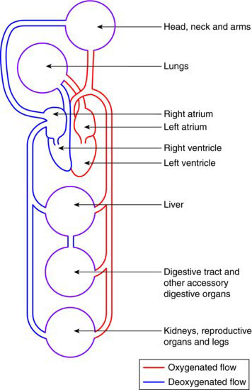
The circulation of the blood throughout the body is represented diagrammatically in Figure 2.1.
Figure 2.1 Body circulation.
THE HEART – GROSS ANATOMY
The heart is a muscular pumping organ situated in the chest cavity, or thorax.
In gross anatomy, it is composed of four chambers – two on the left and two on the right, with no natural communication between the two sides except in the foetus. The ventricles are separated by the interventricular septum.
The left side is concerned with oxygenated blood only, and the right side with deoxygenated blood only.
The two upper chambers are called the atria and the two lower chambers are called the ventricles. The atria receive blood from the body through various veins, while the ventricles deliver blood from within the heart to the body through various arteries.
In effect, the cardiovascular system operates as two interlinked circulatory systems; one taking blood to and from the lungs only – the pulmonary circulation, and the other taking blood to and from the rest of the body – the systemic system.
The gross anatomy of the heart and the passage of blood within it are illustrated in Figure 2.2.
Figure 2.2 Gross anatomy of the heart.
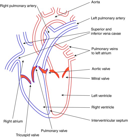
Following the blood flow from the body, through the heart to the lungs and back, and then out to the body again, the sequence is as follows:
- Deoxygenated blood from the body travels to the right atrium in the large veins called the inferior and superior venae cavae
- It is pumped from the right atrium to the right ventricle within the heart through the one-way tricuspid valve, which prevents back flow
- Then from the right ventricle to the lungs in the pulmonary artery – the only artery to carry deoxygenated blood
- The pulmonary valve prevents back flow of blood back into the right ventricle
- The blood is oxygenated in the lungs
- Oxygenated blood travels from the lungs to the left atrium through the pulmonary veins – the only veins to carry oxygenated blood
- It is pumped from the left atrium to the left ventricle within the heart, passing through the one-way mitral valve to prevent back flow
- Finally, it is pumped from the left ventricle out of the heart to the rest of body via the aorta
- This is the largest of the arteries in the body, and back flow is prevented by the aortic valve
- As the aorta leaves the heart, it immediately branches as the coronary arteries, which supply the heart muscle itself with oxygenated blood
- Some of the next branches are the carotid arteries, which supply the head and neck region
THE HEART – MICROSCOPIC ANATOMY
The muscle tissue present in the heart (cardiac muscle) is unique and quite different from that involved in body movement (skeletal muscle) or that around hollow cavities within the body (smooth muscle).
Cardiac muscle has the ability to continue to contract and relax rhythmically, even when it is separated from its nerve supply and is, therefore, not being acted upon by electrical impulses from nerve tissue. In addition, it also does not become tired after a time and cease to contract, like other muscles do. No other muscle tissue has these abilities.
Collectively, the heart wall muscle is known as the myocardium, while the inner surface of the heart is lined by a thin layer of epithelial tissue called the endocardium.
The endocardium is an unbroken layer of tissue that runs into the inner surface lining of the blood vessels, called the vascular endothelium.
THE CARDIAC CYCLE
The heartbeat, or cardiac cycle, occurs about seventy times per minute in the average resting adult human.
It begins in a specialised group of muscle cells on the outer wall of the right atrium, called the sino-atrial node, or ‘pacemaker’.
These specialised cells receive electrical stimulation from two sets of nerves in the brain, one set speeds up the heart rate and the other set slows it down – so the rate of the heart beat is regulated to allow both exercise and rest, as necessary.
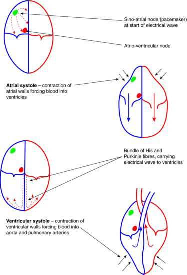
The full cycle occurs as follows:
- Once stimulated at the pacemaker, waves of electrical activity spread quickly across both atria, causing them to contract – this is called atrial systole
- This pumps their blood contents into the ventricles, where it is prevented from back flowing by the tricuspid and mitral valves
- The initial electrical wave stops at this point, after stimulating a second node on the bottom edge of the right atrium, called the atrio-ventricular node
- Electrical impulses from the second node travel in a bundle of specialised cells (bundle of His) lying in the interventricular septum, down to the apex of the heart without spreading across the surfaces of the ventricles
- Once at the apex, the electrical impulse is transmitted from the bottom of the ventricles upwards in specialised fibres (Purkinje fibres), causing ventricular contraction in this same direction – this is called ventricular systole
- The directional nature of the ventricular contraction enables their blood contents to be pumped upwards and out of the heart into the aorta and pulmonary arteries
- Back flow is prevented by the aortic and pulmonary valves
The various valves within the heart are non-muscular structures; they open and close purely due to the fluid pressure differences produced during the contractions of the various heart chambers.
The rhythmic opening and closing of the valves are the heart beat sounds made audible by the use of a stethoscope during clinical examination.
These are often described as ‘lub dup’ – ‘lub’ when the tricuspid and mitral valves simultaneously close, and ‘dup’ when the aortic and pulmonary valves simultaneously close.
The cardiac cycle is shown in Figure 2.3.
Figure 2.3 The cardiac cycle.
BLOOD VESSELS – FUNCTIONS
The three broad types of blood vessel – arteries, veins and capillaries – have each developed to best carry out their particular functions.
Arteries and arterioles (small arteries) specifically transport oxygenated blood from the heart to the body (except the pulmonary artery) and nutrients from the digestive system to the body tissues.
Oxygen is required by all of the body cells to produce energy to work, and nutrients are required for growth and repair.
Veins and venules (small veins) specifically transport deoxygenated blood from the body to the lungs via the heart (except the pulmonary vein) and collect waste products from body tissues to be excreted by the urinary system.
The gaseous waste product of respiration (breathing) is carbon dioxide, while many waste products are produced when nutrients are used by cells and tissues for growth and repair. All these waste products must be eliminated by the body to avoid the development of toxic and acidic conditions, which are damaging to healthy tissues.
Capillaries are the microscopic vessels where these various gaseous and nutrient exchanges occur, lying as networks of vessels that interconnect the arterial and venous sides of the circulatory system.
BLOOD VESSELS – MICROSCOPIC ANATOMY
Arteries and arterioles
These vessels are made up of three layers of tissue, as follows:
- Endothelium – the inner layer of the vessel and in direct contact with the blood
- Smooth muscle – the middle layer of the vessel and surrounds the whole of it with a circular muscle layer, giving the blood vessel strength and the ability to avoid collapse. In the smaller arterioles, more smooth muscle than elastic tissue is present to allow these smaller vessels to regulate the blood flow in their area of supply
- Elastic layer – the outer layer of the vessel and gives it the ability to stretch as a pulse of blood surges through, and then tighten again to ensure the blood flows onwards between heart beats
Those arteries closest to the heart tend to have a thicker elastic layer than the smaller arterioles, so that they are better able to withstand the force of the blood surge as it first leaves the heart, without rupturing.
At the point between the end of ventricular systole and the beginning of the next atrial systole, the blood surge is continued along the length of the arteries to the body tissues by the sequential tightening action of the elastic layer.
The point between the two systoles, when the ventricles are relaxed, is called diastole.
So, the maximum pressure of the blood in the arteries occurs during the peak of ventricular contraction, or systole, while the minimum pressure occurs at the end of ventricular contraction, or diastole.
Hence, blood pressure is recorded as systolic pressure over diastolic pressure, and in a healthy adult at rest it is usually around 120/80.
It is measured as millimetres of mercury, with a sphygmomanometer and a stethoscope.
Capillaries
The arterial blood vessels become smaller and smaller, the further they are from the heart, being called arterioles when they are less than a certain diameter. As they arrive at the tissues they are supplying, they become smaller still and branch repeatedly, until they give rise to capillaries.
Capillaries have the following features:
- Endothelium – made up of just a single layer of cells, so that gases, nutrients and waste products can pass across them easily in an exchange mechanism
- No muscle layer – this would prevent the exchange mechanism from occurring
- No elastic layer – this is not required as the blood flow in these vessels is no longer pulsatile, and again, it would prevent the exchange mechanism from occurring
It is in the capillary beds that the oxygen inspired in the lungs passes out of the circulatory system and into the body tissues to be used to create energy by the cells.
At the same time, carbon dioxide passes from the body tissues into the capillaries to be transported back to the lungs and exhaled. It is formed as the waste product of the cell activities during energy production.
This transfer of the two gases within the capillary beds is called internal respiration.
Nutrients and waste materials are exchanged in a similar manner, by passing out of the capillaries and forming tissue fluid, before being selectively absorbed by the body tissues. Any fluid then not reabsorbed by the capillaries enters a different set of vessels, called the lymph vessels.
The lymph system acts as a safety mechanism, by draining all unwanted materials from the body tissues rather than allowing them to accumulate there, thereby reducing th/>
Stay updated, free dental videos. Join our Telegram channel
VIDEdental - Online dental courses


