CHAPTER 12
Class II, Division 2 Malocclusions
Characteristics of Class II, Division 2 Malocclusions
Class II, division 2 malocclusions are characterized by distoclusion, palatal tipping of two or more maxillary permanent incisors, and lingual tipping of the mandibular incisors. They are differentiated into three types (Fig 12-1). In Type A, all four maxillary permanent incisors are tipped palatally, and the maxillary canines are well aligned in the dental arch. In Type B, the maxillary central permanent incisors are palatally tipped, and the lateral incisors are labially tipped. In Type C, all four maxillary permanent incisors are tipped palatally, and the canines are buccally located, outside the dental arch. However, the essential characteristic of Class II, division 2 malocclusions is the high position of stomion. Stomion is the most anterior point of contact between the upper and lower lips at rest in the midsagittal plane.1 In Class II, division 2 malocclusions, stomion, which is normally located 1 to 3 mm superior to the incisal edges of the maxillary central incisors, is instead at their cervical borders.
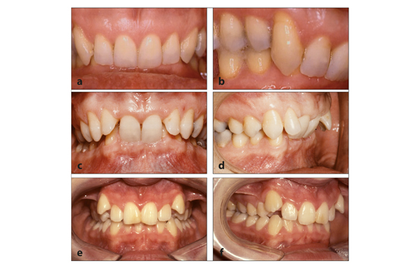
Fig 12-1 Class II, division 2 malocclusions. (a and b) Type A. With excess space in the maxillary dental arch, all four maxillary incisors can tip palatally, and the canines attain a correct position in the dental arch. (c and d) Type B. With limited space, the maxillary central incisors tip palatally, and the lateral incisors tip labially. (e and f) Type C. With marked shortage of space, the four maxillary incisors tip palatally, and the canines emerge buccally outside the dental arch.
Class II, division 2 malocclusions differ from Class II, division 1 malocclusions not only in terms of appearance but also because of etiology. Under highly similar circumstances, either malocclusion can develop (Fig 12-2).
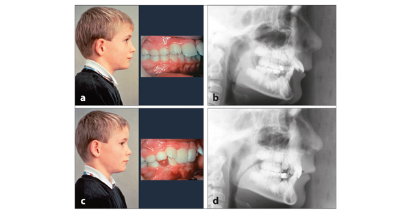
Fig 12-2 Monozygotic twins. (a and b) Class II, division 1 malocclusion. The lower lip is situated behind the maxillary incisors. (c and d) Class II, division 2 malocclusion. The lower lip coverage of the maxillary incisors is excessive. (b and d) The configuration of the facial skeleton is almost identical. (Courtesy of Dr S. Ruf.)
Class II, division 2 malocclusions, and also Class I occlusions with symptoms of Class II, division 2 malocclusions, are called total complete overbite. They are caused by excessive coverage of the maxillary incisors by the lower lip. The risk for this is greater with a short upper lip, a reduced lower anterior facial height, and excess external soft tissues. The lips recede, which increases the force exerted by the lower lip on the maxillary incisors2 (Fig 12-3). However, a total complete overbite can also develop with thin lips if the coverage of the maxillary incisors by the lower lip is excessive.
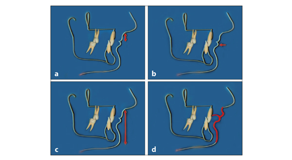
Fig 12-3 Predisposing characteristics for the development of a Class II, division 2 malocclusion. (a and b) A short upper lip. (c) A reduced anterior lower facial height with a horizontally orientated mandibular lower border and a small gonial angle. (d) An excess of labial soft tissues.
Shortly after the emergence of the maxillary central permanent incisors in Class II, division 1 malocclusions, it becomes clear to the casual observer that they are positioned too far to the anterior, particularly if a competent lip seal is lost. The dental characteristics of Class II, division 2 malocclusions show up rather late; initially, there may seem to be no problems.
In the deciduous dentition phase, there is little variation in the labiopalatal inclination of the maxillary incisors. They are oriented nearly perpendicular to the occlusal plane because their successors are situated apical and lingual to their roots.
Initially, Class II, division 2 malocclusions develop as Class II, division 1 malocclusions. In both malocclusions, the mandibular dental arch is positioned too far to the posterior in relation to the maxillary dental arch, ie, there is a distoclusion. The essential difference between the two in the deciduous dentition phase is the height of stomion, which is situated in Class II, division 2 malocclusions at the cervical border of the maxillary central deciduous incisors (Fig 12-4). In Class II, division 1 malocclusions, the lip line is located near the incisal edges of the maxillary deciduous incisors. Through the high position of stomion and the force exerted by the lower lip on the maxillary permanent incisors, two or more of them gradually tip palatally3–8 (Figs 12-5 and 12-6). When the palatally tipped maxillary central incisors start to contact the mandibular central incisors, the latter will tip to the lingual. In a Class I malocclusion with symptoms of a Class II, division 2 malocclusion, this happens at an earlier stage and to a larger extent (Fig 12-7).
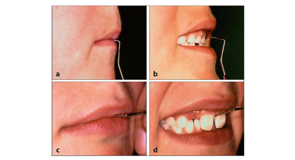
Fig 12-4 The height of stomion in a developing Class II, division 2 malocclusion. (a and b) In the deciduous dentition, the lower lip covers the maxillary incisors. (c and d) After the transition, the lower lip covers an excessive amount of the maxillary permanent incisors.
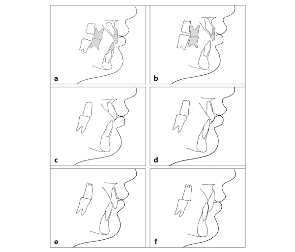
Fig 12-5 Development of a Class II, division 2 malocclusion (compare with Figs 3-4 and 11-5). (a) Stomion is situated near the cervical border of the maxillary deciduous incisors. The overjet is large. (b) The mandibular central permanent incisors emerge normally. The maxillary central permanent incisors start to tip palatally prior to their emergence.9 (c) Due to its high position, the upper lip has only a limited effect on the erupting maxillary central incisors. (d) The latter become more and more influenced by the lower lip, leading to progressive palatal tipping. (e) After sagittal incisal contact is established, the pressure of the lower lip is transferred through the maxillary incisors to the mandibular incisors, which also start to tip lingually. The lower lip covers the maxillary incisors completely. (f) The incisors overerupt in the mandible until they touch the palate and in the maxilla until the gingiva labial to the mandibular incisors is reached.
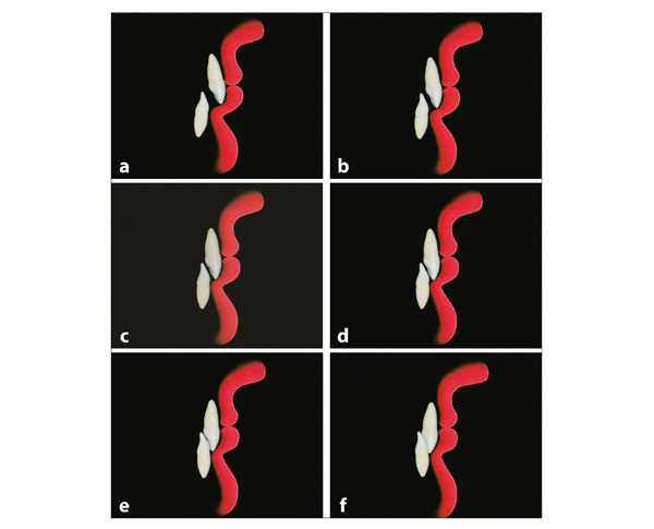
Fig 12-6 Alterations of central permanent incisors in relation to the lips in a Class II, division 2 malocclusion. (a) The mandibular incisors erupt normally. The maxillary incisors have tipped palatally prior to emergence. (b) Thereafter, they continue to tip palatally and overerupt. (c to f ) When contact is established, the mandibular incisors also start to tip lingually. See video clip 33. (Printed from Van der Linden et al10 with permission.)
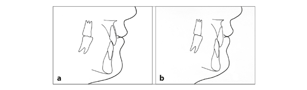
Fig 12-7 Changes in a neutroclusion with symptoms of a Class II, division 2 malocclusion. (a) In a neutroclusion, contact between the incisors is established earlier. (b) The mandibular incisors start to tip lingually sooner; the occurrence and increase of crowding in the mandibular arch also happens earlier.
The difference between a normal transition and the one in a Class II, division 2 malocclusion is demonstrated in Figs 12-8 and 12-9 with longitudinally collected cephalometric radiographs of two children.
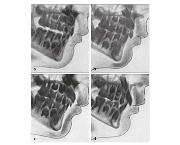
Fig 12-8 Radiographs of normal development in the anterior region. (a) In a normal situation, the maxillary central permanent incisors are formed within the jaw in a labially inclined position. (b) Six months later, the maxillary central permanent incisors have erupted further, and the bone at their labial side has become thinner. (c) Seven months thereafter, their predecessors are still present, and the unemerged permanent incisors have erupted further, without an alteration in angulation, while the bone at the labial side has been resorbed. (d) Eighteen months later, the maxillary central permanent incisors as well as the mandibular incisors have erupted completely and reached occlusal contact. The lower lip lies against the incisal edges of the maxillary incisors and covers only a small portion of their labia/>
Stay updated, free dental videos. Join our Telegram channel
VIDEdental - Online dental courses


