CHAPTER 3
The First Transitional Period: Transition of Incisors and Emergence of First Permanent Molars
Emergence of the First Permanent Molars
Prior to the transition of the incisors, usually at 6 years of age, the first permanent molars emerge: first in the mandible, then in the maxilla. In the preceding years, adequate room was available in the jaws for their development. Continuing posterior growth provided sufficient space for them in the dental arch. They erupt almost perpendicular to the occlusal plane, as the deciduous molars did before them. After their emergence, there is often a surplus of space in the dental arch. Diastemata distal to the second deciduous molars can remain for some time.
The occlusion of the first permanent molars depends on their position in the jaws, the relationship between the mandible and maxilla, and the mesiodistal crown dimensions of the teeth, particularly the mandibular second deciduous molar (Figs 3-1 and 3-2). The width of that tooth varies more than that of any other deciduous tooth. If the mandibular second deciduous molar has a mesiodistal crown dimension that is approximately the same as that of the corresponding maxillary tooth, the terminal plane of the deciduous dentition will have a mesial step. With such a mesial step, the first permanent molars can immediately attain optimal interdigitation (see Figs 3-1a, 3-1b, and 3-2d). However, in most cases the mandibular second deciduous molar is a few millimeters wider than the maxillary second deciduous molar. This results in a flush terminal plane (see Figs 3-1c, 3-1d, 3-2a, and 3-2b). A good mesiodistal occlusion and optimal intercuspation of the first permanent molars cannot occur until the deciduous molars are replaced by the narrower premolars, which have the same crown width. The mandibular first permanent molar then migrates more mesially than the maxillary first molar.

Fig 3-1 Effect of mesiodistal dimensions of second deciduous molars on the occlusion of the first permanent molars. (a) In situations of opposing second deciduous molars with equal mesiodistal crown dimensions, the terminal plane of the deciduous dentition has a mesial step. (b) After emergence, the first permanent molars can arrive at optimal intercuspation immediately. (c) However, in most cases the mandibular second deciduous molar is broader than the opposing maxillary tooth,1 and as a result the terminal plane is flush. (d) Consequently, the first permanent molars erupt with inadequate intercuspation because the cusps are right on top of each other mesiodistally (ie, end-to-end occlusion). However, they do attain an acceptable buccolingual occlusion by means of the cone-funnel mechanism. The maxillary molar is positioned slightly more to the buccal than the opposing mandibular tooth.
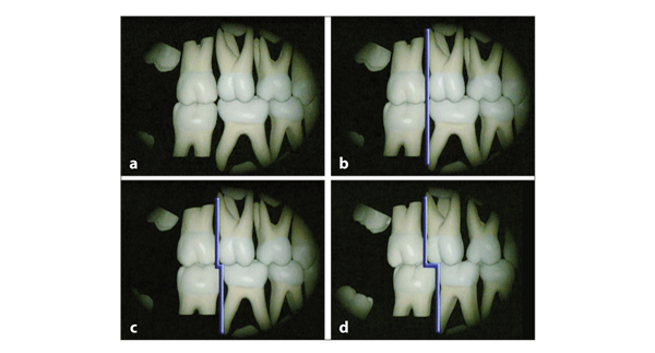
Fig 3-2 Variations in the terminal plane of the deciduous dentition. (a and b) The occlusion of the first permanent molars after their emergence depends on the course of the terminal plane (purple line). A large mandibular second deciduous molar results in end-to-end occlusion of the first permanent molars. (c) A small mandibular second deciduous molar is correlated with a terminal plane with a small mesial step and corresponding suboptimal occlusion of the first permanent molars. (d) In a situation of opposing second deciduous molars with the same mesiodistal crown dimensions, the first permanent molars can immediately attain optimal intercuspation.
Transition of the Incisors
The replacement of deciduous teeth occurs in two phases. In the first transitional period, the incisors are replaced. In the second transitional period, following an interphase of about 1.5 years (the intertransitional period), the deciduous canines and molars are replaced. Marked differences exist between the two transitional periods, mainly due to differences in spatial conditions between the anterior and posterior regions.
Influence of spatial conditions and perioral soft tissues
The crowns of the permanent incisors and canines are broader than those of their predecessors; this difference is greater in the maxilla than in the mandible. Prior to emergence, the tooth buds are located in the region where later their smaller roots will be situated. For the permanent incisors and canines, the circumference of that region is smaller than the arch segment where their incisal margins will be located (Fig 3-3). Again, this difference is greater in the maxilla than in the mandible. In addition, the anterior area is smaller in relation to the sum of the mesiodistal crown dimensions of the six anterior teeth in the maxilla than in the mandible.2
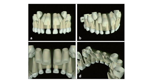
Fig 3-3 Arrangement of maxillary teeth within the surrounding bony structures, some time after the start of eruption of the maxillary central permanent incisors. (a to d) Initially, the permanent incisors are arranged within the jaws in such a way that the available space is used maximally. The median suture does not permit proximity of the central permanent incisors. However, their mesial aspects are parallel and close to the median suture, which makes optimal use of the available space. Consequently, these teeth are distally angulated, erupt in a divergent way, and emerge with a central diastema.
Unlike the deciduous incisors, the permanent incisors erupt in the direction in which they are formed.3,4 Maxillary lateral permanent incisors emerge more labially than the central incisors. After emergence, their labial inclination is reduced through pressure exerted by the lips. In the mandible, the lateral permanent incisors emerge slightly lingual to the dental arch. Subsequently, they migrate labially into alignment with the adjacent teeth through pressure exerted by the tongue. Indeed, the tongue, lips, and cheeks play a major role in the positioning of the teeth. However, local space conditions in the dental arch have the most dominant effect.
The transition of the incisors lasts almost 3 years, measured from the first associated changes in the deciduous dental arch until the complete eruption of all permanent incisors. The loss of the central deciduous incisors and eruption of their successors is illustrated in Fig 3-4. The second deciduous molars and the first permanent molars are also shown. The changes in relation to the tongue and lips from the complete deciduous dentition stage until adulthood are presented in Fig 3-5.
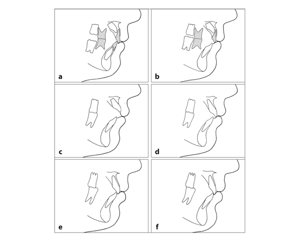
Fig 3-4 Changes in the incisor and molar regions from the complete deciduous dentition stage to adulthood. (a) The deciduous incisors are oriented perpendicular to the occlusal plane. The permanent incisors are located lingually to their predecessors and are labially inclined. (b) First the mandibular first permanent molar emerges, then the mandibular central incisor. The maxillary central permanent incisor emerges some weeks after the loss of its predecessor. (c) In the meantime, the maxillary first permanent molar has emerged. (d) The incisors continue to erupt until contact is reached. (e) The lower lip touches the incisal margin of the maxillary central incisor and covers 1 to 3 mm of its labial surface. (f) Continuing jaw growth and increase of the pressure exerted by the perioral musculature result in a more upright position of the incisors in the adult.
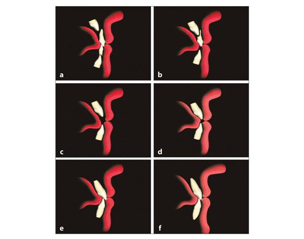
Fig 3-5 Transition of the central incisors in relation to the surrounding soft tissues. (a) The upper lip covers the maxillary deciduous incisors, and the lower lip covers the mandibular incisors as well as a small portion of the maxillary incisors. (b) After emergence, the mandibular permanent incisors contact the lower lip. The incisal margins of the mandibular permanent incisors attain a more labial position than those of the predecessors. (c) This phenomenon occurs to an even greater degree in the maxilla. (d) Prior to and following emergence, the maxillary permanent incisors are more labially inclined than the mandibular incisors. (e) The pressure exerted by the lips leads to a reduction of the labial inclination. The lower lip supports the maxillary incisors vertically together with the mandibular incisors. The roots are completed after emergence in the oral cavity. (f) The skeleton as well as the soft tissues and the nose continue to grow, resulting in a straighter profile and further uprighting of the incisors, more in the maxilla than in the mandible. See video clip 7. (Printed from Van der Linden et al5 with permission.)
Transition of the incisors in a medium anterior section of the apical area
The most common mode of transition of incisors (ie, in a medium anterior section of the apical area) is illustrated in Fig 3-6. The mandibular central deciduous incisors are the first to be lost. Some weeks later their successors emerge, and the overlapping of the lateral incisors reduces. The proximity of the distoincisal corners of the maxillary central permanent incisors to the mesial aspects of the roots of the lateral deciduous incisors causes the latter to migrate distally. This leads to a reduction of the diastemata mesial to the maxillary deciduous canines and to more space for the central permanent incisors. With their further eruption, the deciduous canines migrate distally and buccally.
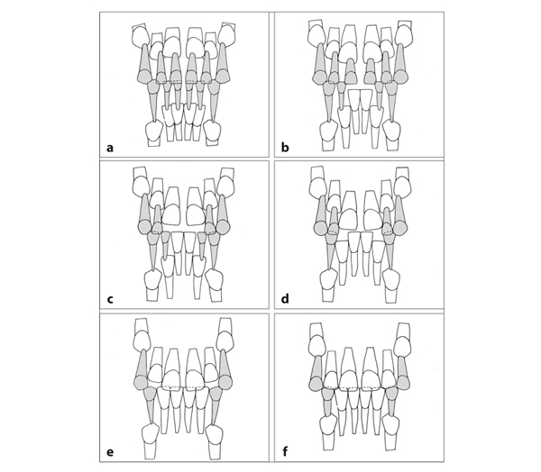
Fig 3-6 The most common mode of transition of incisors. (a) At 5 years of age, the eruption of the permanent incisors has not yet started. (b and c) First the mandibular central deciduous incisors are lost, followed 1 year later by the maxillary central incisors. The distal displacement of the maxillary lateral deciduous incisors is associated with the eruption of the central permanent incisors, which starts a few months earlier. In the mandible, the eruption of the central permanent incisors is not accompanied by migration of deciduous teeth. (d) Shortly before the mandibular lateral permanent incisors emerge, the adjacent deciduous canines move to the distal and buccal. (e) The initially more palatally positioned maxillary lateral permanent incisors erupt in a more labial direction than the central incisors. When their crowns come close to those of the central incisors, the central diastema reduces.6,7 However, that reduction also occurs if this proximity is not reached, as often happens.8,9 With the eruption of the lateral incisors, more room becomes available for the roots, allowing those of the central incisors to move distally. Their mesiodistal angulation alters, and the crowns move toward each other. (f) The maxillary lateral permanent incisors are the last to reach the level of the occlusal plane. There are diastemata in the maxillary anterior region and quite often also in the mandibular region. The roots of the lateral incisors are near the crowns of the permanent canines.
In the mandible, the distoincisal corners of the erupting lateral permanent incisors are in close proximity to the roots of the deciduous canines, which move distally and buccally. Due to the fact that the distal displacement of the deciduous canines occurs in both jaws at about the same time, the increase in intercanine distance usually happens simultaneously.10–15
In both jaws, the lateral permanent incisors emerge about 1 year after the central permanent incisors. The mandibular lateral incisors emerge at about the same time as the maxillary central incisors. Following its emergence, it takes about 8 months for a crown of a maxillary central permanent incisor to become fully exposed.16 (For times of emergence and other statistical data, see chapter 17.)
The modes of transition described above occur when some extra space is needed for adequate eruption and arrangement in the dental arch (ie, a medium anterior section of the apical area). This development is illustrated for the mandible in Fig 3-7, for the maxilla in Fig 3-8, and for both jaws in Fig 3-9.
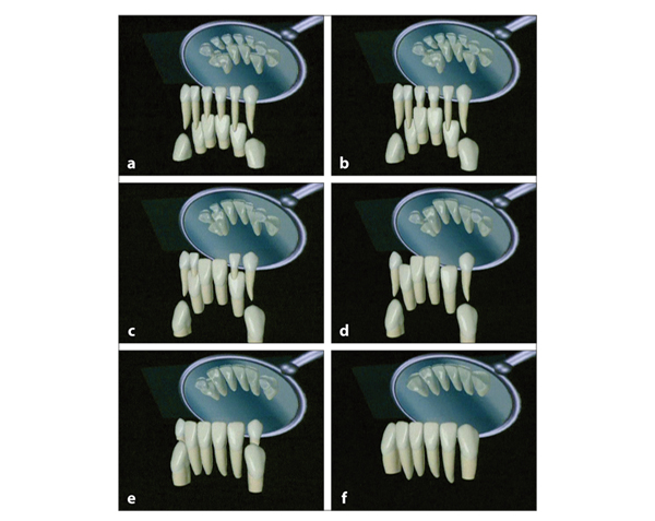
Fig 3-7 Transition of the mandibular incisors in a medium anterior section of the apical area. (a) As indicated, the central permanent incisors are initially located more occlusally than the lateral incisors. They are in close proximity. The roots of the deciduous incisors resorb primarily on the lingual aspect. (b) The central deciduous incisors are lost, and their successors erupt further. (c) They are not in close proximity to the lateral deciduous incisors; as a result, these are not displaced, nor are the deciduous canines. (d) The eruption of the lateral perm/>
Stay updated, free dental videos. Join our Telegram channel
VIDEdental - Online dental courses


