Chapter 1
Basic biology – overview
It is very important for all dental care professionals (DCPs) to have a good underpinning knowledge of the normal structure and function of the human body, and especially of their specialised area – the oral cavity and its surroundings.
This knowledge is essential when trying to understand the subjects of human health, the processes involved in the onset of human disease and the methods available to prevent disease or to treat it when already present.
DEFINITIONS
The scientific discipline that deals with the life processes of living organisms is called biology, and the specific area for this text is human biology.
The particular study of the structures of the human body and their relationships to one another is called human anatomy, and the study of how the body actually functions is called human physiology.
The subject of anatomy covers not only the gross structure of the human body – muscles, bones, organs, and so on – but also the equally important microscopic structures of the cells and tissues themselves that make up these gross structures.
The study of the microscopic structure and function of cells, and the tissues that they form, is called histology.
CELL BIOLOGY
The structures and functions of the human body as a living organism work in a similar way to those of a complicated machine that follows the laws of both chemistry and physics for its operation.
At the most basic level, all the structures of the body are made up of atoms arranged in specific ways that identify them as chemical elements, such as oxygen, hydrogen and carbon. When these different elements combine together they form molecules, such as water and carbon dioxide.
When the molecules themselves combine together in many different ways, they form the basic unit of any living organism – the cell.
The microscopic structure of a basic cell is shown in Figure 1.1 and is composed of the following:
Figure 1.1 Basic structure of the cell.
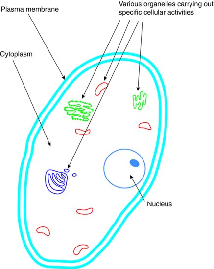
- Plasma membrane – the outer border of the cell itself that holds all of the contents together
- Cytoplasm – the inner fluid of the cell, where all of the internal structures are suspended
- Organelles – specialised structures within the cell, each with their own specific function, such as waste disposal, food production and energy conversion
- Nucleus – the main organelle of all body cells (except red blood cells) that acts as the control centre of the cell by directing the various actions of all of the other organelles, such as energy conversion, growth, food production and waste disposal
It is within the nucleus that the genetic code is stored (usually referred to just as ‘DNA’ by the lay public), so the nucleus controls all of the cell functions and directs its activities.
Most cells are specialised in their actions and perform certain functions within the body. When they are grouped together to carry out these particular functions, they are called tissues.
The main types of tissue found in the body are:
- Muscle tissue – generate forces and produce motion; can be attached to bones to allow the movement of limbs (skeletal muscle), or enclose hollow cavities (smooth muscle) so that they can squeeze around the cavity and remove its contents (such as in the digestive system), or be a third specialised type found only in the heart and called cardiac muscle
- Nervous tissue – initiate and carry electrical impulses along their length to distant areas of the body to effect muscle contractions, gland secretions, and so on
- Epithelial tissue – form the outer layer of the body, as skin, or surround specific groups of tissues to form organs, or line hollow structures within the body, and also separate various areas from each other to avoid the uncontrolled movement of micro-organisms throughout the body
- Connective tissue – connect various parts of the body together to give anchorage and support, such as the connection together of various bones in a joint by tendons and ligaments
- Blood and lymph tissue – are often classed as types of connective tissue, but are unique in existing as cells within a fluid, rather than being a continuous mass of cells like other connective tissues
When several different groups of tissues exist together and carry out a particular function (or functions) they form an organ, such as the heart, the liver, the spleen, and so on.
Organs that have related functions then form integral parts of body systems, such as the digestive system, the respiratory system, and so on.
These will be summarised later.
BASIC TISSUES
The four main basic tissue types will be covered in greater depth to help give a greater understanding of their structure and functions within the oral cavity.
Although separated out in the previous list, blood and lymph will be grouped together with the other connective tissues.
Muscle tissue
This tissue develops from the mesoderm layer of the early embryo, once the developing inner cell cluster has organised into a flat disc at around 3 weeks after fertilisation (see Chapter 6).
All muscle tissue acts by being innervated by the electrical impulses transmitted by nerve cells, causing the contraction (shortening) of the muscle tissue itself. The resultant action produced depends on the surrounding structures that the muscle tissue is connected to.
Muscle tissue can be classified into three groups, depending on their structure, their function and which part of the nervous system activates them:
- Skeletal muscle
- Smooth muscle
- Cardiac muscle
Skeletal muscle has the following characteristics:
- Connected at one or both ends to bones by tendons and ligaments
- Considered as voluntary muscle because they are innervated by somatic nerves (a type of motor nerve), so their contraction can be consciously controlled to produce the voluntary movement of parts of the musculoskeletal system
- Cannot carry out prolonged contraction without tiring, and requiring a rest period
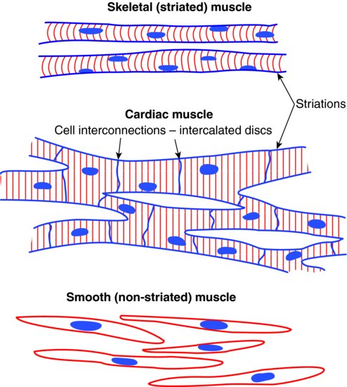
- Microscopically, they are composed of bundles of cell fibres running the length of the tissue, and appear to be striped or striated
- Dentally relevant skeletal muscles include the following:
- Muscles of mastication
- Muscles of facial expression
- The tongue
- Suprahyoid muscles
- Muscles of the pharynx
Smooth muscle has the following characteristics:
- Located in the walls of blood vessels, in hollow organs such as parts of the digestive system, and in glands
- Considered as involuntary muscle because they are innervated by the autonomic nervous system, so their actions cannot be consciously controlled
- Do not tire, as skeletal muscle does
- Microscopically, they are composed of sheets of elongated cells bound in connective tissue, with no striations
- The sheets of cells can run both lengthways and in a circular fashion, so their contraction produces a wave of squeeze-like actions along vessels or in organs and glands
- Unlike skeletal muscle, smooth muscles do not tend to be individually named
- Dentally relevant locations of smooth muscle are:
- All blood vessels
- Salivary glands
- Various areas of the oral cavity, beneath other structures
Cardiac muscle has the following characteristics:
- Specialised type of striated muscle that occurs in the wall of the heart only
- Considered as involuntary muscle because it is innervated by the autonomic nervous system, and cannot be consciously controlled
- Does not tire, otherwise the heart would stop beating and death would occur
- Rate of contraction can be increased or decreased depending on the needs of the body for its supply of oxygenated blood
- Microscopically, the rod-shaped cells run together in a three-dimensional pattern, and interconnect with each other in a grid system so that innervation occurs in a controlled wave across the surface of the heart, producing the heart beat
The microscopic appearance of the three muscle types are illustrated in Figure 1.2.
Figure 1.2 Three types of muscle tissue.
Nervous tissue
This tissue develops from the outer layer of cells of the early embryo, the ectoderm and its specialised sub-layer the neuroectoderm.
The specialised sensory cells of the eyes, the nose, the ears and the taste buds of the tongue are all part of the nervous system, but the basic nerve cell is the neurone.
All neurones function by carrying electrical impulses around the entire body. The electrical impulses can only travel in one direction, so one set of nerve cells transmits impulses from the body to the brain, and another set transmits impulses from the brain to the body.
The neurones of each set of nerve type are arranged in long chains throughout the length of each nerve, interconnecting one with the next neurone in the chain.
Within the brain itself are specialised neurones and their support cells, which are discussed in more detail in Chapter 5.
Nervous tissue making up the peripheral nervous system (that outside the brain and spinal cord) can be simply classified into two groups, depending on the direction of travel of the electrical impulses that they transmit:
- Sensory nerves –carry information from the body to the brain, for analysis and interpretation by the specialised cells within
- Motor nerves –transmit action impulses from the brain to various areas of the body, and can be sub-divided further as follows:
- Somatic nerves – carry impulses to the musculoskeletal system
- Autonomic nerves – carry impulses to blood vessels, organs and glands
A typical neurone is made up of three sections, the cell body and two types of cytoplasmic projection, and is illustrated in Figure 1.3.
Figure 1.3 Neurone.
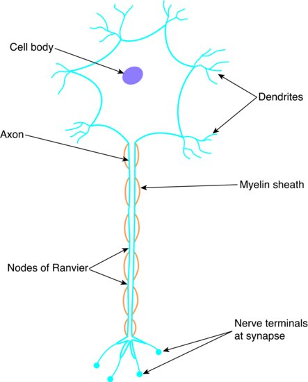
The cell body itself is the support structure for the neurone, and contains all of the usual cellular contents: a nucleus, various organelles, and so on.
Extending from the cell body are various small projections called dendrites, which receive the electrical impulses from the previous neurone and pass them towards the cell body. The cell body itself is not involved in the impulse transmission, and the electrical activity continues on to the other cytoplasmic projection, the axon.
The axon is a much longer projection than the dendrites and is surrounded by its own cell membrane, which is responsible for the continued impulse transmission along the neurone, away from the cell body.
The method of electrical impulse transmission is explained in Chapter 5.
In many nerves throughout the body, the axon also has a fatty covering along its length, interrupted periodically by tiny points of non-coverage called nodes of Ranvier.
In these nerves, the electrical impulse is able to leap-frog along the axon by jumping from node to node, and in this way the impulse is transmitted far more quickly than would otherwise be possible.
The point at which one neurone connects with another, or with the tissue that it is to innervate, is called the synapse. At this point, the electrical impulse is further transmitted by chemicals called neurotransmitters.
Epithelial tissue
This tissue develops from all three layers of cells in the early embryo – the ectoderm, the mesoderm and the endoderm – and the layer of origin determines the final tissue and its function in the body.
So, a simple overview is as follows:
- Ectoderm origin – develop into the skin and the inner linings of hollow body organs and systems, such as the digestive tract and the lungs
- Mesoderm origin – develop into the epithelial sheaths surrounding the heart, the lungs and the abdominal cavity, and referred to as the mesothelium
- Endoderm origin – develop into the inner linings of blood vessels, and referred to as the vascular endothelium
/>
Stay updated, free dental videos. Join our Telegram channel
VIDEdental - Online dental courses


