Fig. 1.1
Anterior of human skull
The human nose is built from a “bridge” that is formed by a pair of nasal bones that meet at the midline. Maxillae, or commonly known as the upper jaw, are a union of two bones at the hard palate. This bone forms part of the orbital floor, part of the nasal cavity lateral walls and floor as well as most of the hard palate [4]. Every bone of the face has articulation with maxilla except the mandible, which is the lower jawbone. The hard palate is formed by the palatine processes of the maxilla and the horizontal plates of the palatine bones. A large maxillary sinus soft tissue is contained in each maxilla and it is unfilled into the nasal cavity. The maxilla also consists of sockets for the upper teeth positioning, called the alveolar process. In addition, there is an opening in the maxilla inferior to the orbit known as infraorbital foramen. It works as a passage of the infraorbital nerve and blood vessels and a branch of the maxillary division of the trigeminal (V) nerve. The other foramen that exists in maxilla is the incisive foramen. It is located in posterior to the incisor teeth. The inferior orbital fissure can be found between the greater wing of the sphenoid and the maxilla [2].
Zygomatic bones or commonly called cheekbones are another paired bone of the skull. It is located at the upper and lateral part of the face and forms the prominence of the cheek, part of the lateral wall and floor of each orbit and parts of the temporal and infratemporal fossa. This zygoma represents four processes, which are the frontosphenoidal, orbital, maxillary and temporal process, with four borders. The temporal process of zygoma has an articulation with the zygomatic process of the temporal bone to form the zygomatic arch [4]. Zygoma is free from bone resorption or regeneration compared to other surrounding bones like maxillae [5]. Based on literature studies, zygoma is appropriate to be used as anchorage for implant placement. A long fixture can be installed within the bone starting from alveolar process in maxillae together with ordinary fixtures. The fixture or implant could be used as anchorage for epithesis, prosthesis and obturators [2].
The two lacrimal bones are the smallest bones of the face. These bones located at the posterior and lateral to the nasal bones and they form part of medial wall of the orbits. Palatine bones are paired bones with L-shaped that form the posterior portion of the hard palate, part of the lateral wall and floor of the nasal cavity as well as a small portion of the orbital floors. The vomer is the bone on the floor of the nasal cavity. It is also communicates superiorly with the perpendicular plate of the ethmoid bone and inferiorly with maxillae and palatine bones along the midline [2, 4]. The mandible or also known as lower jawbone is the largest and strongest facial bones. The bone has curvy shape, horizontal part, the body and two vertical parts. The two vertical parts or called as rami or in singular is ramus, has a condylar process, which articulates with mandibular fossa and articular tubercle of the temporal bone to form temporomandibular joint (TMJ). Similar with maxillae, the mandible also has the alveolar process (sockets) for the lower teeth configuration.
1.2 Dental Anatomy
The tooth structure can be divided into two main parts—the crown and the root as illustrated in Fig. 1.2. The crown can be classified into two portions, the anatomical and the clinical crown. The anatomical crown is part of crown that is found in gingival soft tissue whilst the clinical crown is part of crown that is exposed. Teeth consist of several tissue structures like enamel, dentine, cementum and the dental pulp [4]. Enamel is the most outer surface of a tooth and it is the hardest tissue in human body [1]. It acts as a protection to the dentine, which is softer than enamel but harder than bone tissues [6]. It is a living tissue and must be protected during surgical operation. The teeth root is covered by bonelike tissue called the cementum, which is a thin layer tissue with the main function of providing teeth anchorage to the bony wall in periodontium. The periodontium or periodontal ligament provides a gap between the cementum and the alveolar bone. It has a height of approximately 2 mm. The dental pulp contains blood vessels and nerves and it cannot be separated from the dentine either anatomically or functionally [6]. Among functions of the pulp are to form the dentine, provide sensation to the teeth and respond to irritation. The major parts normally involve in most dental practice are teeth, dental arches and periodontal tissue.
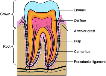
Fig. 1.2
Structure of tooth
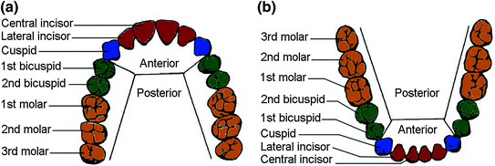
Primary teeth in children consist of 20 units and the number increases to 28–32 and will be permanent for adults [1, 4]. The 20 units primary teeth are equally distributed in terms of number for the maxilla and mandible. This is also true for the permanent teeth where a maximum of 16 units are located in the maxilla and mandible respectively. The teeth configuration can be divided into four quadrants, two quadrants for the maxilla and the other two quadrants for the mandible. Each quadrant has two incisors, one cuspid (canine), two bicuspids (premolar) and three molars as shown in Fig. 1.3a and b [3, 4]. Incisors can be classified into central and lateral incisor. The central incisors are located on both sides of the midline of the arch central location. Next to the central incisor is the lateral incisor. Both incisors act like scissors during mastication [7]. The teeth located after the incisors are the canine followed by the two premolars (the first and second premolars). The canine has a sharp cusp that effectively tears and shred foods while the premolars have two pointed projections for similar function plus an additional ability to crush food. The most posterior teeth are the three molars; first molar, second molar and third molar. The molars are the strongest and most useful tooth with a main function to grind food into tiny pieces [4, 7].
Stay updated, free dental videos. Join our Telegram channel
VIDEdental - Online dental courses


