The individual tooth positions and inclinations will be described in relationship to a horizontal reference plane. This reference plane is recorded from the patient and transferred to an articulator. Many different techniques and articulator components exist, and are beyond the scope of this chapter. However, a very practical technique that can be applied to most articulation systems will be presented.
Esthetic reference plane
The occlusal plane by definition is the average plane established by the incisal and occlusal surfaces of the teeth or in the case of the edentulous patient, the surface of wax occlusion rims contoured to guide in the arrangement of denture teeth.7 The occlusal plane can be viewed in both the frontal and sagittal planes. The incisal surfaces, when viewed in the frontal plane, should have a pleasing parallelism to the patient’s facial features (interpupillary line, eyebrows, hairline, etc.) (Fig. 11-2).
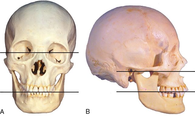
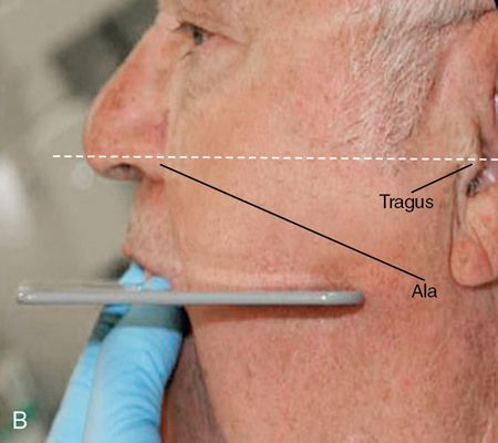
The posterior occlusal surfaces when viewed in the sagittal plane should, on average, correspond to an imaginary plane established by the inferior border of the ala of the nose (or the average between the two) and the superior border of the tragus of each ear (Camper’s plane).7 Although the accuracy of Camper’s plane as representative of the occlusal plane is arguable, it is adequate for this proposed technique. This ideal plane will be referred to as the esthetic reference plane (ERP) (see Fig. 11-2). Fabricating a restoration that is parallel with the patient’s ERP (especially when viewed in the frontal plane) is critical to the esthetic success of the restoration. Restorations that deviate from this horizontal plane are typically the result of improper or inadequate communication with the dental laboratory technician. Therefore it is imperative that the patient’s ERP be recorded by the clinician and then communicated to the technician. The simplest and least expensive tool that facilitates this evaluation is the Biteplane (Ivoclar Vivadent) or Fox Plane (Dentsply International).
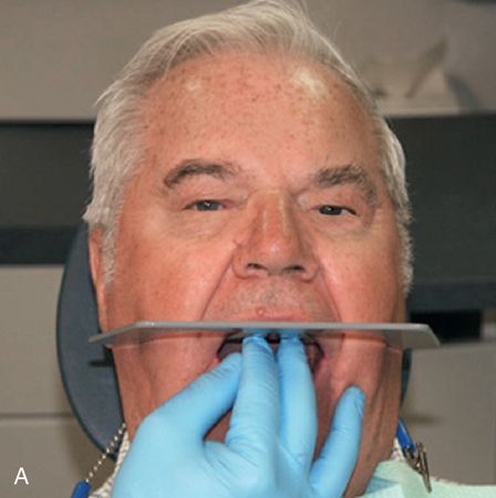
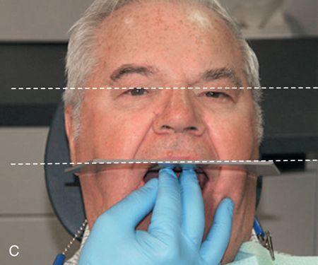
The Biteplane rests on the surfaces of the maxillary teeth or in the case of complete dentures, the maxillary wax rim. The external wings of the Biteplane help to amplify the visual relationship of the patient’s maxillary incisal/occlusal plane to their ERP (Fig. 11-3 A,B).
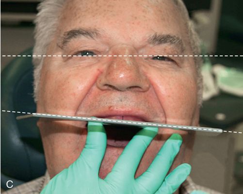
If the incisal and/or occlusal surfaces are not in harmony with the patient’s ERP (Fig. 11-3 C), the wax rim is corrected by either removing or adding wax.
Once the wax occlusion rim is determined to be acceptable, it then becomes the prescription to the dental lab concerning the midline, length, and facial position of the maxillary anterior teeth as well as being representative of the patient’s ERP.
Relating the ERP to the articulator
There are several ways to communicate this information to the dental laboratory. In this technique a flat mounting table (Ivoclar Vivadent) (Fig. 11-4 A,B) is used. The table has premarked average value positions that will facilitate the positioning and mounting of the maxillary cast to an average value relationship to the condyles of the articulator.
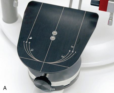
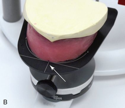
For the edentulous patient the wax rim is positioned so that the incisal edge rests on the premarked position and the midline (obtained from the patient) corresponds to the midline of the table (see Fig. 11-4 A)
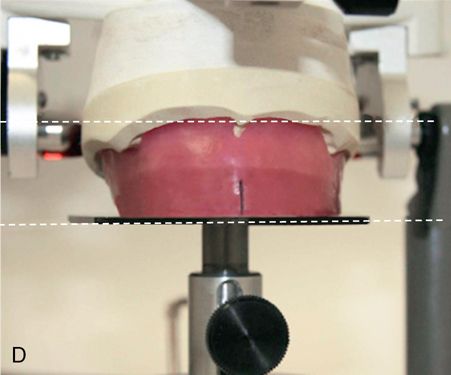
The maxillary cast is then mounted to the upper member of the articulator and the surface of the flat mounting table is now representative of the patient’s ERP (Fig. 11-4 C,D). The surface of the table also serves as a plane of reference for tooth positioning.
Tooth position options
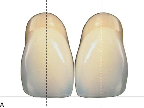
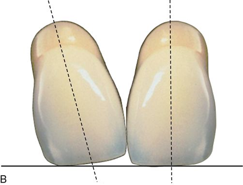
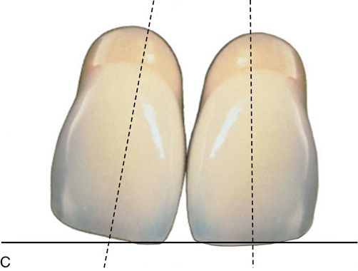
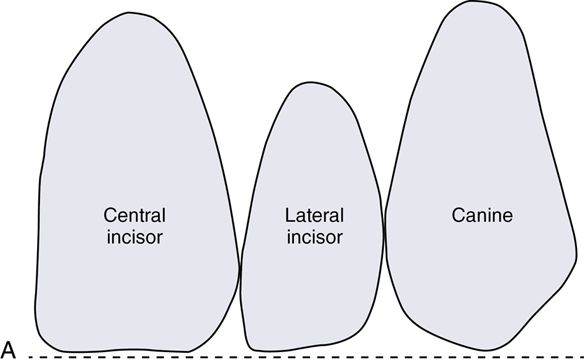
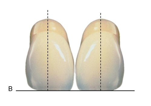
There are four positions in which to set anterior teeth: mesiodistal inclination, labiopalatal inclination, rotation, and length (see Fig. 11-1). Mesiodistal inclination is viewed from the frontal plane. When the incisal edges are flush to the ERP the mesiodistal inclination is typically perpendicular to the ERP and is considered to have no inclination (Fig. 11-5 A). If the tooth is inclined toward the mesial, it is described as having mesial inclination (Fig. 11-5 B) If the tooth is inclined toward the distal, it is described as having mesial inclination (Fig. 11-5 C).
Labiopalatal inclination.
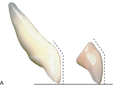
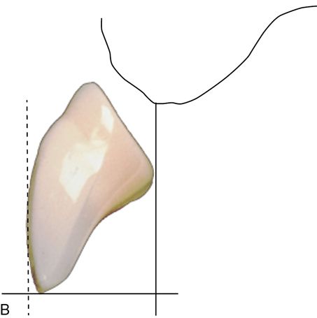
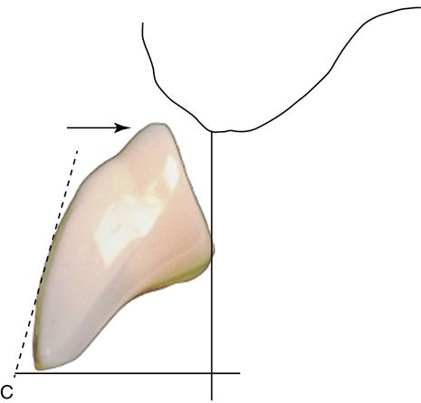
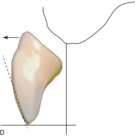
Labiopalatal inclination is viewed from the sagittal plane. The facial surface of maxillary anterior teeth is divided into two planes. The incisal plane follows the coronal two thirds of the tooth and the cervical plane follows the apical third of the tooth (Fig. 11-6 A). If the incisal plane is perpendicular to the ERP, the labiopalatal inclination is described as having no inclination (Fig. 11-6 B). If the incisal plane of the tooth is inclined toward the labial, it is described as having labial inclination (Fig. 11-6 C). If the incisal plane is inclined toward the palate, it is described as palatal inclination (Fig. 11-6 D).
Rotation.
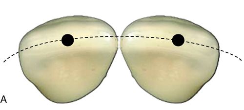
Rotation is viewed from a perspective perpendicular to the incisal edges of anterior teeth and the cusp tips of posterior teeth and visualizing a center axis line of rotation. If the incisal edge follows the semicircular arch-form, the tooth will automatically have a slight rotation, which positions the mesial of the tooth slightly facially. This will be referred to as follow-the-arch form (Fig. 11-7 A).
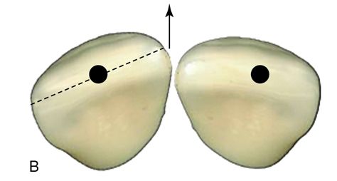
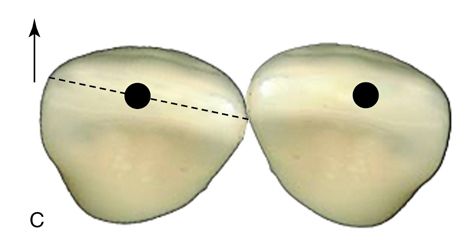
If the mesial surface of the tooth is rotated facially around the center axis to a degree, which is outside the arch form (i.e., the mesial surface is more prominent) it is considered to have a mesial flare and will accentuate the mesial highlight of the tooth. This will give the tooth a smaller and softer appearance (Fig. 11-7 B).
If the distal surface of the tooth is rotated facially around the center axis to a degree that is outside the arch form (i.e., the distal surface is more prominent) it is considered to have a distal flare, which gives the tooth a larger and more bold appearance and the highlight is positioned more toward the center of the tooth (Fig. 11-7 C).
Height.
The height of a tooth is determined by whether or not it contacts the flat mounting table and is described by the number of millimeters the tooth is elevated from the surface of the table (i.e., the measured space between the incisal edge and the surface of the table) (Fig. 11-8).
Rules of dental esthetics
Figure 11-9 outlines the four tooth positions previously described and applies them to each maxillary anterior tooth individually. The various esthetic changes that occur are described and illustrated in the rest of the chapter.
Stay updated, free dental videos. Join our Telegram channel
VIDEdental - Online dental courses


