8: The Permanent Canines: Maxillary and Mandibular

8 The Permanent Canines: Maxillary and Mandibular

The maxillary and mandibular canines bear a close resemblance to each other, and their functions are closely related. The four canines are placed at the “corners” of the mouth; each one is the third tooth from the median line, right and left, in the maxilla and mandible. They are commonly referred to as the cornerstone of the dental arches.1 They are the longest teeth in the mouth; the crowns are usually as long as those of the maxillary central incisors, and the single roots are longer than those of any of the other teeth. The middle labial lobes have been highly developed incisally into strong, well-formed cusps. Crowns and roots are markedly convex on most surfaces. The shape and position of the canines contribute to the guidance of the teeth into the intercuspal position by “canine guidance.”2

The shape of the crowns, with their single pointed cusps, their locations in the mouth, and the extra anchorage furnished by the long, strongly developed roots, makes these canines resemble those of the carnivore. This resemblance to the prehensile teeth of the carnivore gives rise to the term canine.

Because of the labiolingual thickness of crown and root and the anchorage in the alveolar process of the jaws, these teeth are perhaps the most stable in the mouth. The crown portions of the canines are shaped in a manner that promotes cleanliness. This self-cleansing quality, along with the efficient anchorage in the jaws, tends to preserve these teeth throughout life. When teeth are lost, the canines are usually the last ones to go. They are very valuable teeth, when considered either as units of the natural dental arches or as possible assistants in stabilizing replacements of lost teeth in prosthetic procedures.

Both maxillary and mandibular canines have another quality that must not be overlooked: the positions and forms of these teeth and their anchorage in the bone, along with the bone ridge over the labial portions of the roots, called the canine eminence, have a cosmetic value. They help form a foundation that ensures normal facial expression at the corners of the mouth. Loss of all of these teeth makes it extremely difficult, if not impossible, to make replacements that restore that natural appearance of the face for any length of time. It would therefore be difficult to place a value on the canines, and their importance is manifested by their efficiency in function, stability, and aid in maintaining natural facial expression.

In function, the canines support the incisors and premolars, since they are located between these groups. The canine crowns have some characteristics of functional form, which bears a resemblance to incisor form and also to the premolar form.

Maxillary Canine

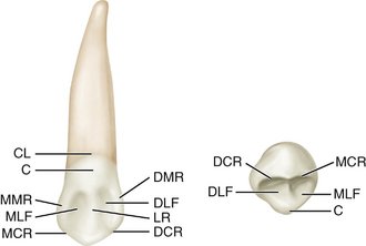
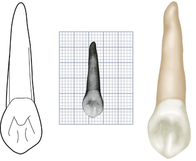
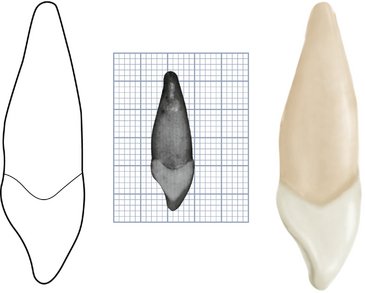
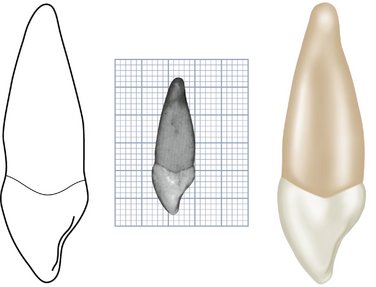
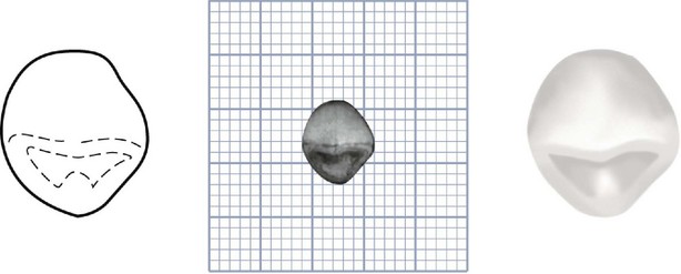
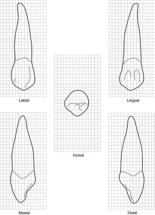
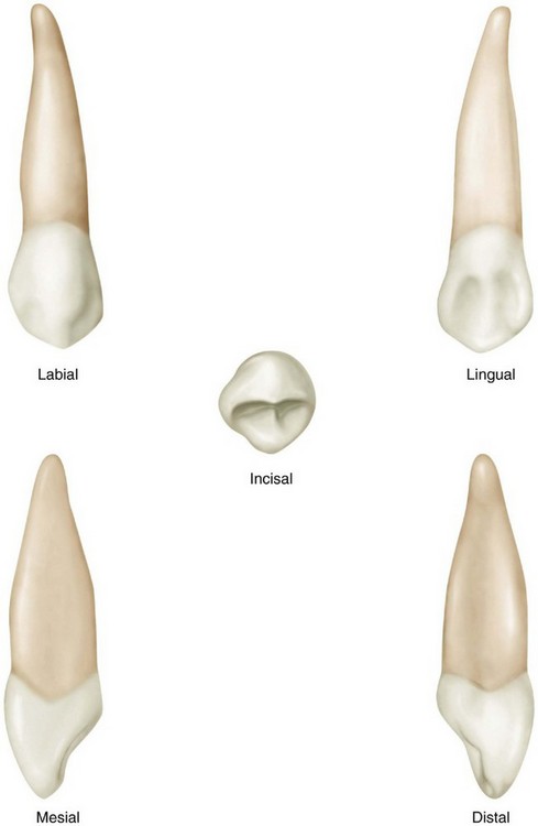
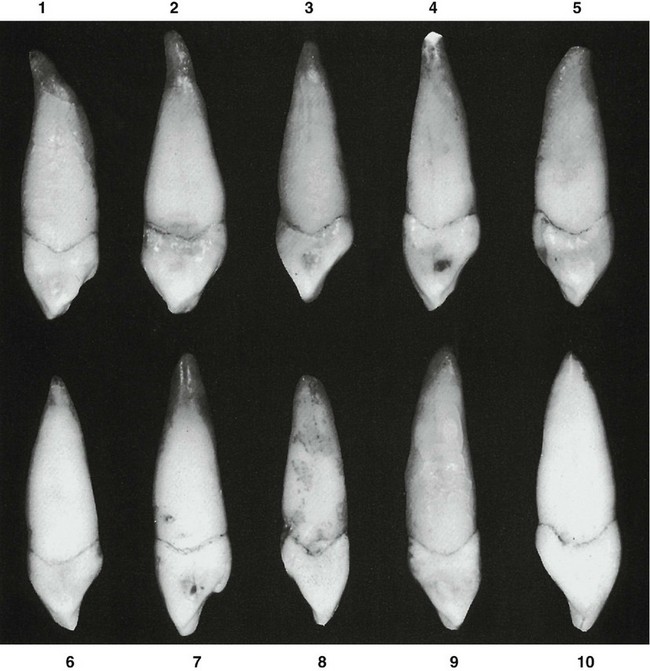
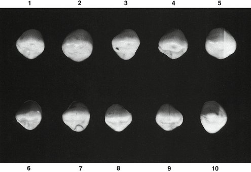
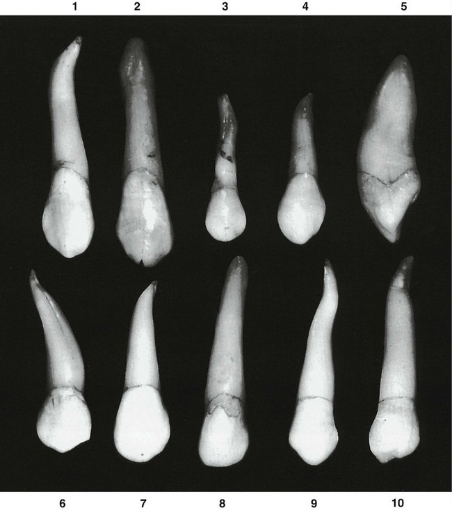
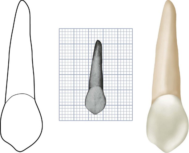
Figures 8-1 through 8-12 illustrate the maxillary canine in various aspects. The outline of the labial or lingual aspect of the maxillary canine is a series of curves or arcs except for the angle made by the tip of the cusp. This cusp has a mesial incisal ridge and a distal incisal ridge.

The mesial half of the crown makes contact with the lateral incisor, and the distal half contacts the first premolar. Therefore the contact areas of the maxillary canine are at different levels cervicoincisally.

From a labial view, the mesial half of the crown resembles a portion of an incisor, whereas the distal half resembles a portion of a premolar. This tooth seems to be a compromise in the change from anterior to posterior teeth in the dental arch.

It is apparent that the construction of this tooth has reinforcement, labiolingually, to offset directional lines of the force brought against it when in use. The incisional (incising) portion is thicker labiolingually than that of either the maxillary central or the lateral incisor.

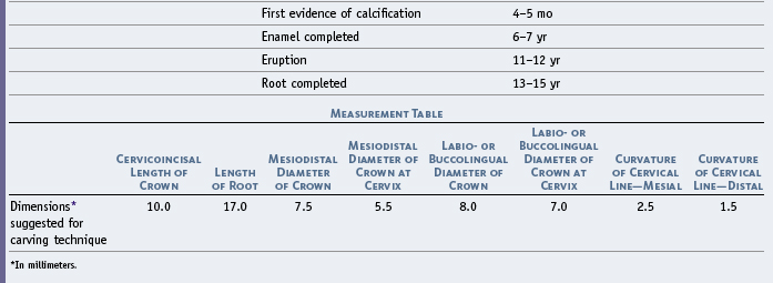
The labiolingual measurement of the crown is about 1 mm greater than that of the maxillary central incisor (Table 8-1). The mesiodistal measurement is approximately 1 mm less.

The cingulum shows greater development than that of the central incisor.

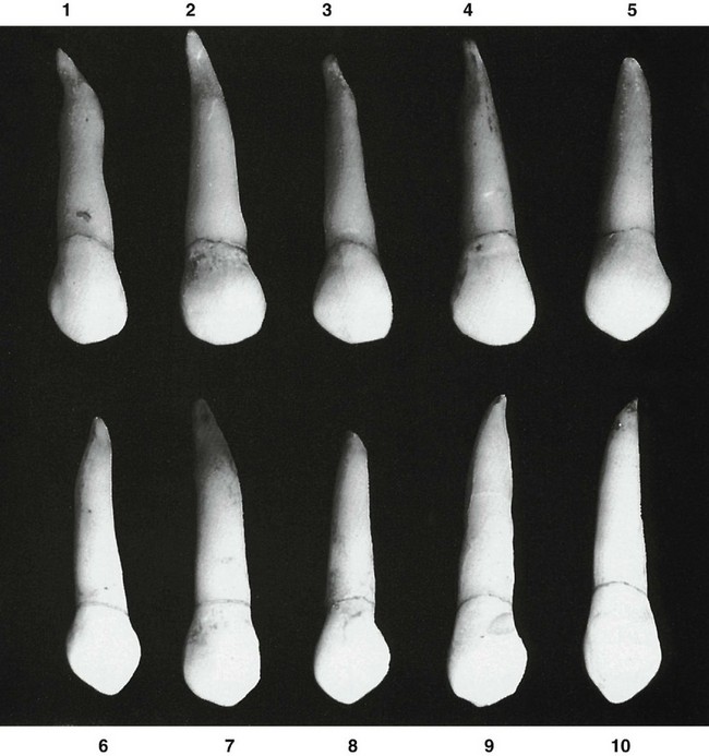
The root of the maxillary canine is usually the longest of any root with the possible exception of that of the mandibular canine, which may be as long at times. The root is thick labiolingually, with developmental depressions mesially and distally that help furnish the secure anchorage this tooth has in the maxilla. Uncommon variations are shown in Figure 8-12.

DETAILED DESCRIPTION OF THE MAXILLARY CANINE FROM ALL ASPECTS

Labial Aspect

From the labial aspect, the crown and root are narrower mesiodistally than those of the maxillary central incisor. The difference is about 1 mm in most mouths. The cervical line labially is convex, with the convexity toward the root portion (see Figures 8-2, 8-7, 8-8, and 8-9).

Mesially, the outline of the crown may be convex from the cervix to the center of the mesial contact area, or the crown may exhibit a slight concavity above the contact area from the labial aspect. The center of the contact area mesially is approximately at the junction of middle and incisal thirds of the crown.

Distally, the outline of the crown is usually concave between the cervical line and the distal contact area. The distal contact area is usually at the center of the middle third of the crown. The two levels of contact areas mesially and distally should be noted (see Figure 5-7, B and C).

Unless the crown has been worn unevenly, the cusp tip is on a line with the center of the root. The cusp has a mesial slope and a distal slope, the mesial slope being the shorter of the two. Both slopes show a tendency toward concavity before wear has taken place (see Figure 8-9, 5 and 6). These depressions are developmental in character.

The labial surface of the crown is smooth, with no developmental lines of note except shallow depressions mesially and distally, dividing the three labial lobes. The middle labial lobe shows much greater development than the other lobes. This produces a ridge on the labial surface of the crown. A line drawn over the crest of this ridge, from the cervical line to the tip of the cusp, is a curved one inclined mesially at its center. All areas mesial to the crest of this ridge exhibit convexity except for insignificant developmental lines in the enamel. Distally to the labial ridge (see incisal aspect), a tendency exists toward concavity at the cervical third of the crown, although convexity is noted elsewhere in all areas approaching/>

Stay updated, free dental videos. Join our Telegram channel

Jan 9, 2015 | Posted by in Occlusion | Comments Off on 8: The Permanent Canines: Maxillary and Mandibular

VIDEdental - Online dental courses
Get VIDEdental app for watching clinical videos