CHAPTER 7
The Adult Dentition
This chapter deals with the complete permanent dentition. As way of introduction, some aspects of development are mentioned, although most of them have been covered in the previous chapters in another context. Finally, the worn dentition of an elderly individual is shown.
Tooth Positions
After all permanent teeth except the third molars have emerged at 12 or 13 years of age, alterations in tooth positions still take place. The roots of the permanent incisors, particularly the maxillary incisors, move distally. After emergence of the third molars, the roots of the maxillary permanent molars spread throughout the available space. If the third molars are absent, this probably occurs earlier. The roots of the second molars move distally, and their angulation alters. Ultimately, all maxillary molars are mesially angulated. The distal margins of the maxillary first molars become positioned more occlusally than the adjacent mesial margins of the second molars.1 All maxillary teeth except the second premolars are mesially angulated. The maxillary posterior teeth are inclined slightly buccally; the incisors are inclined labially. Mesiodistally, the mandibular premolars are oriented perpendicular to the occlusal plane. The lateral incisors, canines, and molars are mesially angulated. The molars are inclined lingually; the incisors and canines are inclined labially. The premolars are the turning point between the two inclinations (Figs 7-1 to 7-4).
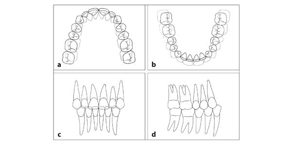
Fig 7-1 Permanent dentition. (a) The maxillary teeth are arranged in an arch without diastemata. They occlude slightly outside of the mandibular ones. There is a small overjet. (b) The mandibular teeth are in contact with each other and form an arch on which the maxillary teeth fit. All mandibular teeth have occlusal contact with two maxillary teeth, with the exception of the mandibular central incisors. (c and d) The roots of the maxillary lateral incisors are directed distally, as are those of the central incisors, although to a slightly lesser degree. The roots of the mandibular lateral incisors are slightly distally angulated. The premolars intercuspate like gears.
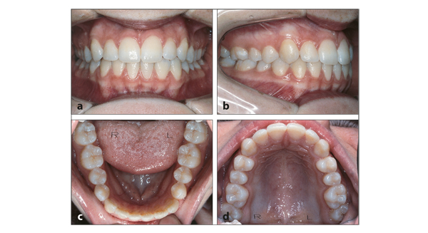
Fig 7-2 Ideal permanent dentition. (a to d) In this case, all criteria of an ideal position and occlusion of teeth are met, including the course of the gingival line. The texture and color of the gingiva and the height of the smile line are also optimal. As a result of the availability of sophisticated orthodontic appliances, an ideal arrangement can be reached in many cases.
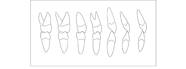
Fig 7-3 Inclination of permanent teeth. The maxillary incisors and canines are more labially inclined than the mandibular incisors and canines. The lateral incisors are more labially inclined than the central incisors, particularly in the maxilla. In the maxilla, the premolars are buccally inclined. In the mandible, their buccolingual orientation is perpendicular to the occlusal plane. The maxillary molars are buccally inclined; the mandibular molars are lingually inclined. Note that the occlusal contacts in this illustration deviate from reality. The distal sides of the teeth are drawn, not cross sections of the points where opposing teeth meet.

Fig 7-4 Angulation of permanent teeth and the relationship between their length and the level where their formation started (shaded). All teeth are mesially angulated, except both mandibular premolars and the maxillary second premolar. These teeth are oriented perpendicular to the occlusal plane. In both the maxilla and the mandible, the canines are formed at the farthest distance from the occlusal plane and are ultimately the longest teeth. In the maxilla, the lateral incisor, which is formed closer than the central incisor to the occlusal plane, is the shortest maxillary incisor. In the mandible, the situation is reversed.
Factors Affecting the Arrangement of the Teeth
In every population, a large diversity exists in the position and occlusion of permanent teeth. The angulation and inclination of teeth vary considerably, even in normal occlusion2 (see chapter 17, Table 17-30). The same also applies to the shape and relationship of the jaws and to the influence of functional factors. The position of the apices depends on the morphology of the jaws, the spatial conditions within the jaws, and their sagittal and transverse relationships. The position of the crowns and their inclinations are further determined by the occlusion and the pressures exerted by the tongue, lips, and cheeks. In that regard, the dimensions and volume of the soft tissues are of importance, but their position in unstrained conditions is even more essential. Furthermore, the shape and size of the crowns, the tooth-supporting tissues, and the forces generated by the functioning dentition affect the position of the individual teeth. In this context, habits such as thumb sucking, chewing of tobacco and coca leaves, bruxism, and tooth grinding must be considered. In addition, many factors alter slightly with age, including the morphology of the jaws and the craniofacial skeleton and also the tone, texture, and form of the soft tissue. Most of the aspects mentioned above are clarified further in chapter 9.
Criteria for an Ideal Dentition
Indeed, the arrangement of the teeth depends on many factors, and an ideal occlusion is quite rare. In most cases, there are some minor shortcomings. A variety of malocclusions, from mild to severe, are found in every population (see chapters 10 to 15). The criteria for an ideal dentition are not limited to specific norms for tooth positions and occlusion but also include requirements for the course of the gingival line. In the maxilla, the clinical crowns of the lateral incisors should be shorter than those of the central incisors, which in turn should be shorter than the canine crowns. The course of the gingival line corresponds with these differences in clinical crown heights, ie, it is lower at the lateral incisors than at the central incisors and again higher at the canines. In these aspects, symmetry is important.
In the mandibular anterior region, the h/>
Stay updated, free dental videos. Join our Telegram channel
VIDEdental - Online dental courses


