7
Cervical Spine and Soft Tissues of the Neck
Anatomy—Cervical Spine and Soft Tissues of the Neck
This section will highlight anatomical landmarks of the cervical spine, specifically the cervical vertebrae.
As cone beam CT scans are not made to differentiate soft tissue, this chapter will not identify specific soft tissue anatomy. It will look at more general areas, many of which are only identifiable when using a scan with a large field of view.
Axial
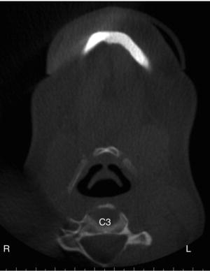
The axial figures start from the superior aspect of the cranium moving inferiorly (Figures 7.1–7.4).
Figure 7.1. Axial view at the level of the inferior aspect of the maxillary sinuses showing the anterior arch of C1 (AA-C1) and the odontoid process of C2 (OP-C2).
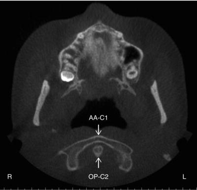
Figure 7.2. Axial view at the level of the mandibular foramina showing the odontoid process of C2 (OP-C2) and posterior arch of C1 (PA-C1).
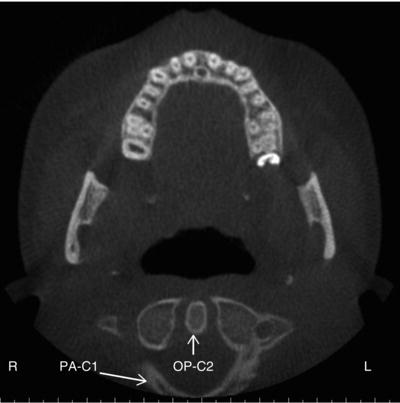
Figure 7.3. Axial view at the level of mandibular teeth at the cemento-enamel junction showing the entire arch of C2.
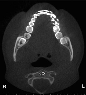
Figure 7.4. Axial view at the level of the hyoid bone showing the entire arch of C3.
Coronal
The coronal figures start from the anterior aspect of the face moving posteriorly (Figures 7.5–7.8).
Figure 7.5. Coronal view at the aspect of the auditory canal showing the anterior arch of C1 (AP-C1) and portions of the body of C2.
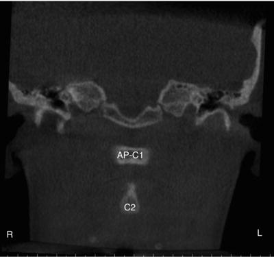
Figure 7.6. Coronal view at the aspect of the mastoid air cells showing cervical vertebrae C1, C2, and C3.
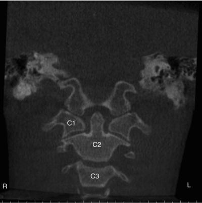
Figure 7.7. Coronal view at aspect of the posterior cranial base showing C1, C2, C3, and portions of C4.
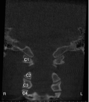
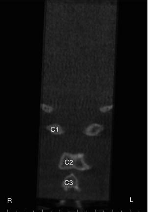
Figure 7.8. Coronal view showing C1, C2, and C3.
Sagittal
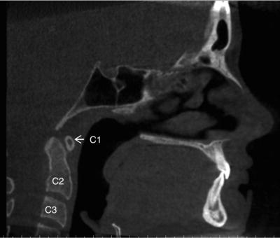
The sagittal figures start from the lateral aspect of the cranium moving medially (Figures 7.9–7.10).
Figure 7.9. Sagittal view at the lateral aspect of the maxillary posterior teeth showing cervical vertebrae C1, C2, C3, and portions of C4.
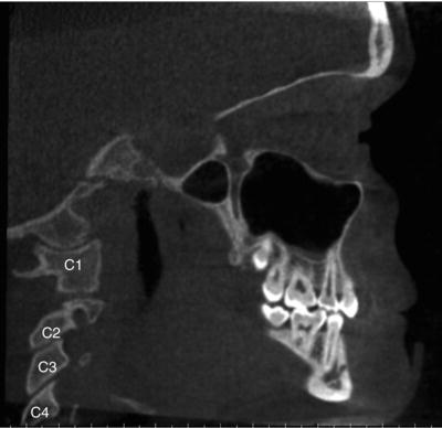
Figure 7.10. Sagittal view on the midline showing cervical vertebrae C1, C2, and C3.
Incidental Findings
Developmental
There are many variations of developmental anomalies that may present in the cervical vertebrae. This book cannot encompass them all and will list some of the most commonly occurring ones. The developmental anomalies are listed in order of most superior cervical vertebrae moving inferiorly.
Clefts (C1)
Definition/Clinical Characteristics
They are incomplete fusions of the ossification centers of either the anterior or posterior arch of C1 (atlas) or a combination of both. The fusion of the anterior arch occurs by the age of 10 and the posterior arch completes fusion between the ages of 3 or 4. Posterior arch clefts have an incidence of 4% of all adults with 97% of these clefts occurring in the midline. Anterior arch clefts are less common, occurring in only 0.1% of all adults. Cervical vertebral clefts occur more commonly in patients with cleft lip, cleft palate, or both.
Radiographic Description
It appears as a well-defined radiolucent line or band of the anterior or posterior arch of C1. There may be multiple radiolucent lines (thee or more) visible in the anterior arch (Figure 7.11). The posterior arch rarely presents with more than one radiolucent line. The bony borders of the arch and ossification centers are corticated. When both anterior and posterior clefts are noted, the appropriate descriptive term is split atlas. This is best visualized on axial and coronal views (Figures 7.12 and 7.13).
Differential Interpretation
Radiolucent line(s) in the anterior or posterior arch of C1 that are not corticated means one would need to consider the possibility of a fracture. A fracture typically presents with swelling combined with a history of trauma to the area.
Figure 7.11. (a) Coronal view showing three anterior arch clefts (black arrows) in a 5 year 11 month old; (b) Axial view showing three anterior arch clefts (black arrows) in a 5 year 11 month old.
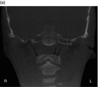
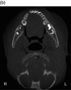
Figure 7.12. (a) Axial and (b) coronal views showing a single anterior arch cleft (black arrow) in a 20 year old.
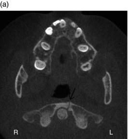
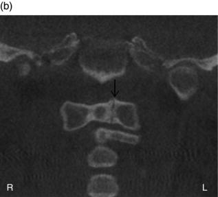
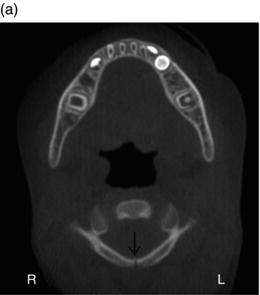
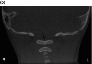
Figure 7.13. (a) Axial and (b) coronal views showing a single posterior arch cleft (black arrow).
Treatment/Recommendations
There is no recommended treatment or further imaging necessary for this finding.
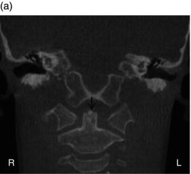
Os Terminale (C2)
Definition/Clinical Characteristics
There are three ossification centers that develop to form the body of C2 (axis). Two form the body and one forms the tip of the odontoid process. The ossification center that forms the tip of the odontoid process is referred to as os terminale. It is typically evident by the age of 3 and fuses with the body of C2 by the age of 12. It has been reported in 1.3%–9.9% of children.
Radiographic Description
Os terminale presents as a well-defined radiopaque entity at the tip of the body of C2. The body of C2 has a V-shaped notch on thesuperior aspect where the ossification center is located on coronal views (Figure 7.14). There is a complete to incomplete radiolucent line separating the ossification center from the remainder of the body of C2. This is best visualized on coronal and sagittal views (Figure 7.15).
Figure 7.14. Coronal views (a,b) showing os terminale at the superior aspect of the odontoid process of C2 (black arrow).
Stay updated, free dental videos. Join our Telegram channel
VIDEdental - Online dental courses


