5
Cranial Skull Base
Anatomy
This section will highlight anatomical landmarks of the cranial skull base and the bones they are a part of. Many of these landmarks are only identifiable when using a scan with a large field of view.
Axial
The axial figures (Figures 5.1–5.5) start from the superior aspect of the cranium moving inferiorly. The following anatomy is readily identifiable on axial views (Table 5.1).
Figure 5.1. Axial slice at the level superior to the orbits showing the frontal sinus (star).
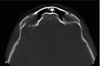
Figure 5.2. Axial slice at the level of the superior aspect of the orbits showing the crista galli (black arrow).
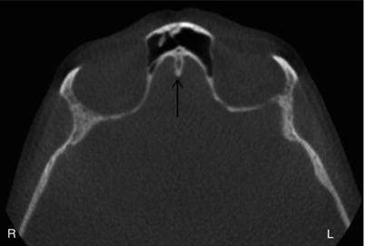
Figure 5.3. Axial slice at the level of the midorbits showing the following sphenoid bone anatomy: the anterior clinoid process (A), sella turcica (S), and posterior clinoid process (P).
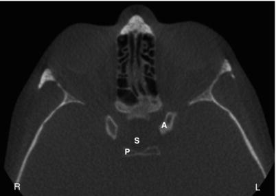
Figure 5.4. Axial slice at the level of the inferior orbit/superior maxillary sinus showing the foramen lacerum (FL), carotid canal (CC), and petrous ridge (PR).
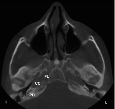
Figure 5.5. Axial slice at the level of the midmaxilla showing the mastoid process with mastoid air cells (MAC), styloid process (SP), clivus/occipital bone (C), foramen magnum (FM), and pterygoid process (PP).
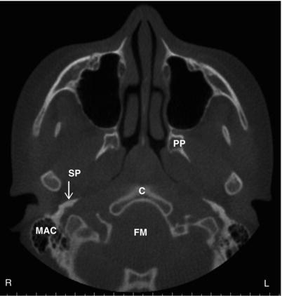
Table 5.1. Anatomical landmarks identifiable on axial views with corresponding figures.
| Bone | Anatomical landmark | Figures visible on |
| Frontal | Frontal sinus | 5.1 |
| Ethmoid | Crista galli | 5.2 |
| Sphenoid | Anterior clinoid process Sella turcica Posterior clinoid process Foramen lacerum Pterygoid process |
5.3 5.3 5.3 5.4 5.5 |
| Temporal | Carotid canal Petrous ridge Mastoid process Styloid process |
5.4 5.4 5.5 5.5 |
| Occipital | Clivus / occipital bone Foramen magnum |
5.5 5.5 |
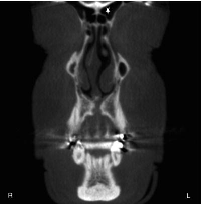
Figure 5.6. Coronal slice at the anterior aspect of the nasal cavity showing the frontal sinus (star).
Coronal
The coronal figures (Figures 5.6–5.11) start from the anterior aspect of the face moving posteriorly. The following anatomy is readily identifiable on coronal views (Table 5.2).
Figure 5.7. Coronal slice at the lateral aspect of the orbits showing the cribriform plate (white arrow) and crista galli (black arrow).
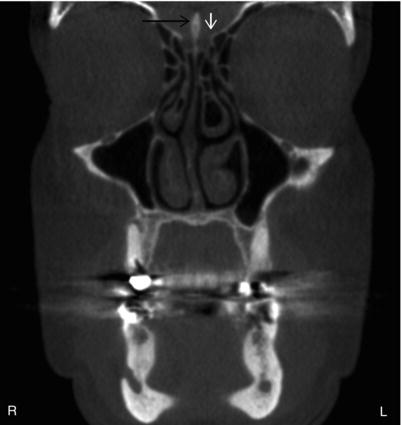
Figure 5.8. Coronal slice at the aspect of the coronoid process showing the pterygoid process (PP), vidian canal (VC), foramen rotundum (FR), and anterior clinoid process (A).
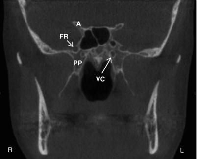
Figure 5.9. Coronal slice at the posterior aspect of the ramus showing the posterior clinoid process (P) and foramen lacerum (FL).
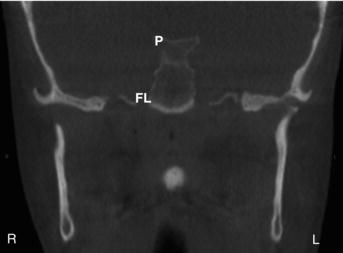
Figure 5.10. Coronal slice at the aspect of the mastoid process showing the mastoid process with mastoid air cells (MAC) and depression of the sigmoid sinus (SS).
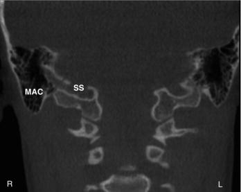
Figure 5.11. Coronal slice at the posteriormost aspect showing the jugular foramen (JF) and petrous ridge (PR).
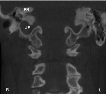
Table 5.2. Anatomical landmarks identifiable on coronal views with corresponding figures.
| Bone | Anatomical landmark | Figures visible on |
| Frontal | Frontal sinus | 5.6 |
| Ethmoid | Cribriform plate Crista galli |
5.7 5.7 |
| Sphenoid | Anterior clinoid process Posterior clinoid process Foramen lacerum Foramen rotundum Pterygoid process Vidian canal |
5.8 5.9 5.9 5.8 5.8 5.8 |
| Temporal | Petrous ridge Mastoid process Sigmoid sinus depression |
5.11 5.10 5.10 |
| Occipital | Jugular foramen | 5.11 |
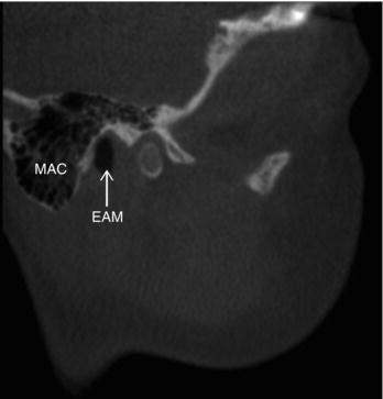
Figure 5.12. Sagittal slice at the lateral aspect of the mastoid process showing the mastoid process with mastoid air cells (MAC) of the temporal bone is evident along with the external auditory meatus (EAM).
Sagittal
The sagittal figures (Figures 5.12–5.15) start fro/>
Stay updated, free dental videos. Join our Telegram channel
VIDEdental - Online dental courses


