6
Soft Tissue of the Brain and Orbits
Anatomy—Soft Tissue of the Brain and Orbits
As cone beam CT scans are not made to differentiate soft tissue, this chapter will not identify specific soft tissue anatomy but more general areas. This anatomy is identifiable when using a scan with a large field of view.
Axial
The axial figures start from the superior aspect of the cranium moving inferiorly (Figures 6.1–6.5).
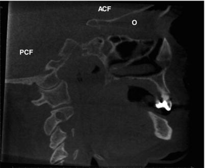
Figure 6.1. Axial slice at the level of the superior aspect of the orbits (O) showing the anterior cranial fossa (ACF).
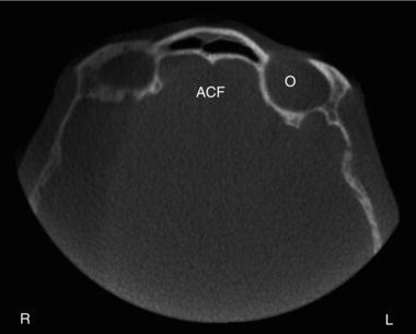
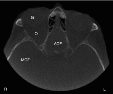
Figure 6.2. Axial slice at the level of the midorbits (O) showing the anterior cranial fossa (ACF), middle cranial fossa (MCF), and globe of the eye (G).
Coronal
The coronal figures start from the anterior aspect of the face moving posteriorly (Figures 6.6–6.11).
Sagittal
The sagittal figures start from the lateral aspect of the cranium moving medially (Figures 6.12–6.15).
Figure 6.3. Axial slice at the level of the inferior orbit (O) and superior maxillary sinus showing the middle cranial fossa (MCF) and posterior cranial fossa (PCF).
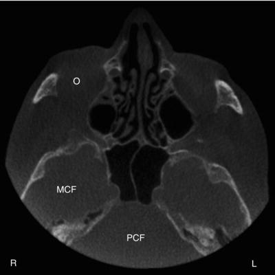
Figure 6.4. Axial slice at the level of the midmaxillary sinus showing the posterior cranial fossa (PCF).
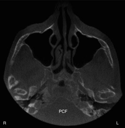
Figure 6.5. Axial slice at the level of the inferior maxillary sinus showing the posterior cranial fossa (PCF).
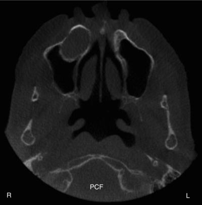
Figure 6.6. Coronal slice at the anterior aspect of the maxillary sinuses showing the orbits (O).
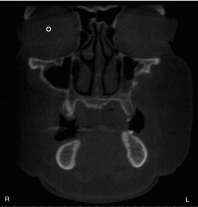
Figure 6.7. Coronal slice at the midmaxillary sinus and midorbits (O) showing the anterior cranial fossa (ACF).
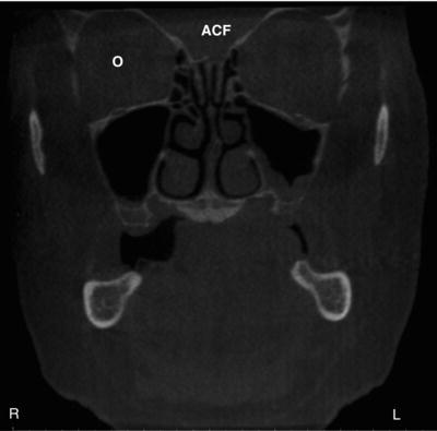
Figure 6.8. Coronal slice at the posterior aspect of the orbits (O) showing the anterior cranial fossa (ACF).
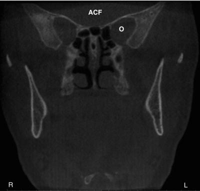
Figure 6.9. Coronal slice at the posterior aspect of the ramus showing the middle cranial fossa (MCF).
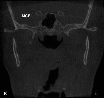
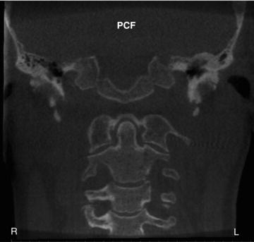
Figure 6.10. Coronal slice at the midcervical vertebrae showing the posterior cranial fossa (PCF).
Incidental Findings—Soft Tissue of the Brain
Incidental Calcifications
Choroid Plexus Calcification
Definition/Clinical Characteristics
Choroid plexus calcifications are common physiologic calcifications that increase with age. They occur in approximately 12% to 43.3% of the population.
Figure 6.11. Coronal slice at the posterior aspect of the cervical vertebrae showing the posterior cranial fossa (PCF).
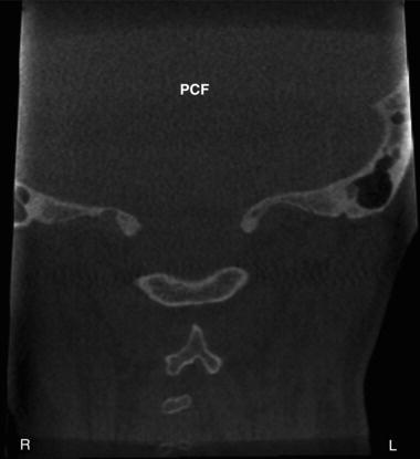
Figure 6.12. Sagittal slice at the lateral aspect of the mastoid process showing the middle cranial fossa (MCF).
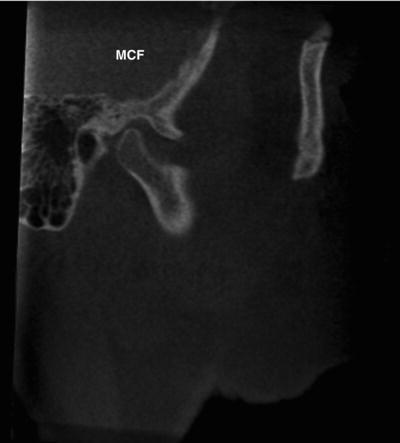
Figure 6.13. Sagittal slice at the lateral aspect of the angle of the mandible showing the middle cranial fossa (MCF), posterior cranial fossa (PCF), and globe of the eye (G).
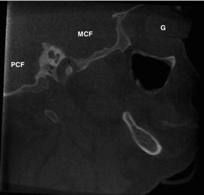
Figure 6.14. Sagittal slice at the lateral aspect of the nasal cavity showing the anterior cranial fossa (ACF), posterior cranial fossa (PCF), and orbits (O).
Radiographic Description
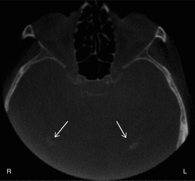
Choroid plexus calcification is noted in the posterior aspect of the soft tissue of the brain. It is either unilateral or bilateral. It appears as diffuse radiopaque entities lateral to the midline (Figure 6.16). They are commonly visualized on axial and coronal views (Figure 6.17).
Figure 6.15. Sagittal slice on the midline showing the anterior cranial fossa (ACF) and posterior cranial fossa (PCF).
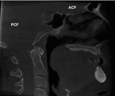
Figure 6.16. Axial slice at level of midorbits showing bilateral choroid plexus calcifications (arrows) lateral to the midline.
Differential Interpretation
There are no differential interpretations based on the location of this finding.
Treatment/Recommendations
No further treatment or imaging is recommended.
Figure 6.17. (a) Coronal slice at aspect of posterior cervical vertebrae showing bilateral choroid plexus calcifications (arrows) lateral to the midline; (b) Coronal slice at posterior to cervical vertebrae showing bilateral choroid plexus calcifications (arrows) lateral to the midline near the superior aspect of the field of view.
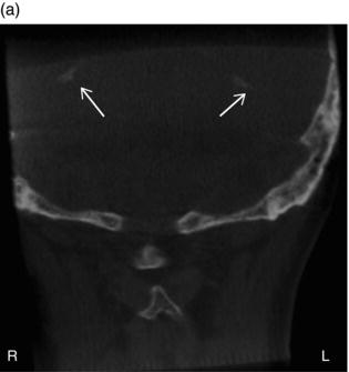
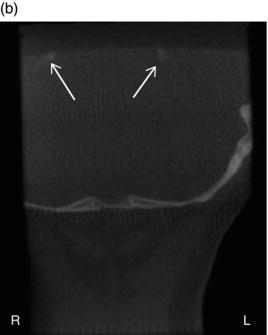
Figure 6.18. Axial slice at the level of the superior midorbital region showing dural calcifications (arrow).
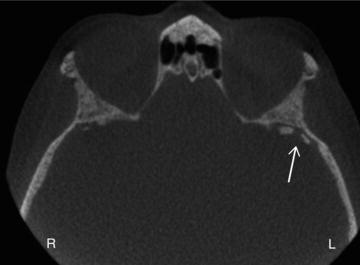
Dural Calcifications
Definition/Clinical Characteristics
Dural calcifications are age-related calcifications found in the dura of the brain. They occur in approximately 10% of the elderly population.
Radiographic Description
Dural calcifications may occur near the cranium or in the midline. They appear as either linear radiopaque entities in the midline to radiopaque masses near the cranium (Figure 6.18). They are visualized on all views (axial, coronal, and sagittal; Figure 6.19).
Figure 6.19. (a) Coronal and (b) sagittal slices at/>
Stay updated, free dental videos. Join our Telegram channel
VIDEdental - Online dental courses


