6
Regional differences in the oral mucosa
6.1 STRUCTURAL VARIATIONS IN DIFFERENT REGIONS
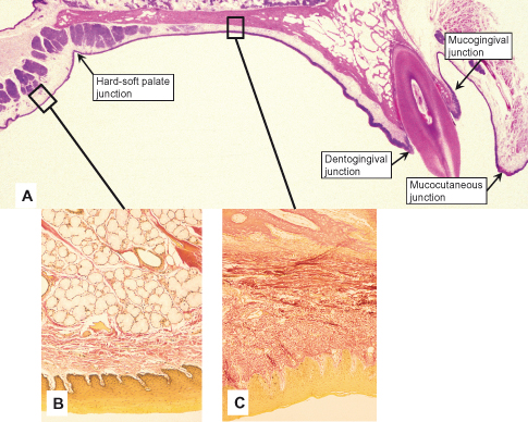
The range of regional variation seen in oral mucosa is, if we exclude appendages such as nails and hair, even greater than that seen in skin. This range includes differences not only in the composition of the lamina propria, form of the interface between epithelium and connective tissue, and type of surface epithelium, but also in the nature of the submucosa and how the mucosa is attached to underlying structures. These regional differences are considered to represent functional adaptations, and the mucosa is consequently divided into three main types: masticatory, lining, and specialized. The regions occupied by each type have been illustrated in Figure 2.1. A summary of the mucosal organization within the various anatomic regions is given in Table 6.1, and a more detailed description follows below. Finally, a brief account is given of junctions between different types of mucosa that are of morphologic interest and clinical importance (Fig. 6.1A).
Table 6.1 Structure of the mucosa in different regions of the oral cavity.
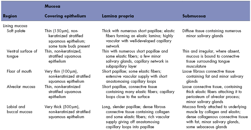
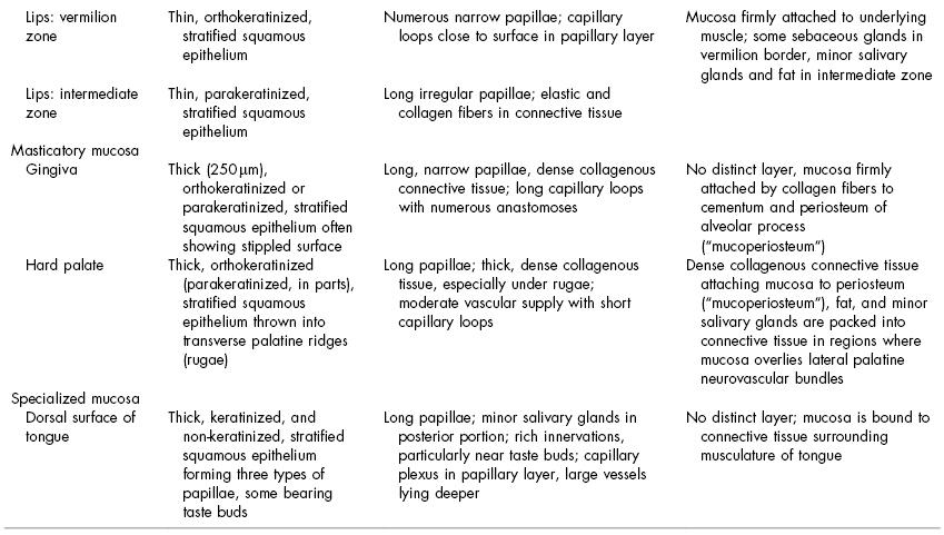
Figure 6.1 Junctions in the oral mucosa. (A) Histologic section cut sagittally through the mandible and anterior vestibule showing the junction between the lining mucosa (enlarged in B) of the soft palate and the masticatory mucosa of the hard palate (enlarged in C), the interface between the masticatory mucosa of the gingiva and the enamel of the tooth (dentogingival junction), the interface between the attached gingiva and the alveolar mucosa (mucogingival junction), and the interface between the skin and the oral mucosa (mucocutaneous junction).
6.1.1 Masticatory Mucosa
Masticatory mucosa covers those areas of the oral cavity such as the hard palate and gingiva (Fig. 6.1A) that are exposed to compressive and shear forces and to abrasion during the mastication of food (see Fig. 2.1). The dorsum of the tongue has the same functional role as other masticatory mucosa but, because of its specialized structure, it is considered separately.
The epithelium of masticatory mucosa is moderately thick. It is frequently orthokeratinized, although normally there are parakeratinized areas of the gingiva and occasionally of the palate. Both types of epithelial surface are inextensible and are well adapted to withstanding abrasion. The junction between epithelium and underlying lamina propria is convoluted, and the numerous elongated papillae probably provide good mechanical attachment and prevent the epithelium from being stripped off under shear force. The lamina propria is thick, containing a dense network of collagen fibers in the form of large, closely packed bundles (Fig. 6.1C). They follow a direct course between anchoring points so that there is relatively little slack and the tissue does not yield on impact, enabling the mucosa to resist heavy loading.
Masticatory mucosa covers immobile structures (e.g., the hard palate and alveolar processes) and is firmly bound to them either directly by the attachment of lamina propria to the periosteum of underlying bone—such as in mucoperiosteum—or indirectly by a fibrous submucosa. In the lateral regions of the hard palate, this fibrous submucosa is interspersed with areas of fat and glandular tissue that cushion the mucosa against mechanical loads and protect the underlying nerves and blood vessels of the palate.
As already mentioned in Chapter 2, the firmness of masticatory mucosa ensures that it does not gape after surgical incisions (see Fig. 2.4B) and rarely requires suturing. For the same reason, injections of local anesthetic into these areas are difficult and are often painful, as is any swelling arising from inflammation.
6.1.2 Lining Mucosa
The oral mucosa covering the underside of the tongue, inside of the lips, cheeks, floor of the mouth, and alveolar processes as far as the gingiva is mobile. These regions, together with the soft palate, are classified as lining mucosa (see Fig. 2.1).
The epithelium of lining mucosa is thicker than that of masticatory mucosa, sometimes exceeding 500 µm in the cheek, and is non-keratinized. The surface is thus flexible and is able to withstand stretching. The interface with connective tissue is relatively smooth, although slender connective tissue papillae often penetrate into the epithelium.
The lamina propria is generally thicker than in masticatory mucosa and contains fewer collagen fibers, which follow a more irregular course between anchoring points (Fig. 6.1B). Thus, the mucosa can be stretched to a certain extent before these fibers become taut and limit further distention. Associated with the collagen fibers are elastic fibers that tend to control the extensibility of the mucosa. Where lining mucosa covers muscle, it is attached by a mixture of collagen and elastic fibers. As the mucosa becomes slack during masticatory movements, the elastic fibers retract the mucosa toward the muscle and so prevent it from bulging between the teeth and being bitten.
The alveolar mucosa and the floor of the mouth mucosa are loosely attached to the underlying structures by a thick submucosa. In contrast, mucosa of the underside of the tongue is firmly bound to the underlying muscle. The soft palate is flexible but not highly mobile, and its mucosa is separated from the loose and highly glandular submucosa by a layer of elastic fibers.
The tendency for lining mucosa to be flexible, with a loose and often elastic submucosa, means that surgical incisions frequently require sutures for closure (see Fig. 2.4A). Injections into such regions are easy because there is ready dispersion of fluid in the loose connective tissue; however, infections also spread rapidly. The lining mucosa of the floor of the mouth and underside of tongue is thinner and is more permeable than the other regions (see Chapter 8) and, consequently, has been used for systemic delivery of pharmaceutical agents and local delivery of antigens to elicit mucosal immune response.
6.1.3 Specialized Mucosa
The mucosa of the dorsal surface of the tongue is unlike that anywhere else in the oral cavity because, although covered by what is functionally a masticatory mucosa, it is also a highly extensible lining and, in addition, has different types of lingual papillae. Some of them possess a mechanical function, whereas others bear taste buds and therefore have a sensory function.
The mucous membrane of the tongue is composed of two parts, with different embryologic origins, and is divided by the V-shaped groove, the sulcus terminalis (terminal groove). The anterior two-thirds of the tongue, where the mucosa is derived from the first pharyngeal arch, is often called the body, and the posterior third, where the mucosa is derived from the third pharyngeal arch, the base. The mucosa covering the base of the tongue contains extensive nodules of lymphoid tissue, the lingual tonsils.
Fungiform Papillae
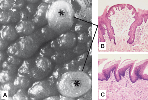
The anterior portion of the tongue bears the fungiform (“funguslike”) and filiform (“hairlike”) papillae (Fig. 6.2A). Single fungiform papillae are scattered between the numerous filiform papillae at the tip of the tongue (Fig. 6.2B). They are smooth, round structures that appear red because of their highly vascular connective tissue core, visible through a thin, non-keratinized covering epithelium. Taste buds are normally present in the epithelium on the superior surface.
Figure 6.2 Lingual papillae from the dorsal surface of the tongue. (A) Fungiform papillae are interspersed among numerous filiform papillae on the anterior of the tongue, two of which are evident (asterisks). (B,C) Histologic sections of the papillae in (A) show that the epithelium of the fungiform papilla is thinly keratinized or non-keratinized (B) and that of the filiform papillae is keratinized (C).
Filiform Papillae
Filiform papillae cover the entire anterior part of the tongue and consist of cone-shaped structures, each with a core of connective tissue, covered by a thick keratinized epithelium (Fig. 6.2C). Together, they form a tough, abrasive surface that is involved in compressing and breaking food when the tongue is apposed to the hard palate. Thus, the dorsal mucosa of the tongue functions as a masticatory mucosa. Buildup of keratin results in elongation of the filiform papillae in some patients. The dorsum of the tongue then has a “hairy” appearance called hairy tongue.
The tongue is highly extensible, with changes in its shape accommodated by the regions of non-keratinized, flexible epithelium between the filiform papillae.
Foliate Papillae
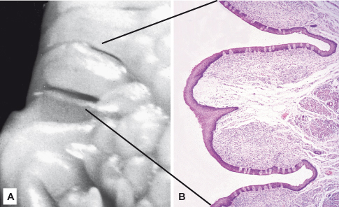
Foliate (“leaflike”) papillae are sometimes present on the lateral margins of the posterior part of the tongue, although they are more frequently seen in mammals other than humans (Fig. 6.3A). These pink-colored papillae consist of 4– 11 parallel ridges that alternate with deep grooves in the mucosa, and numerous taste buds are present in the epithelium of the lateral walls of the ridges (Fig. 6.3B).
Figure 6.3 (A) Ridge-shaped foliate papillae are another type of lingual papillae located laterally on the tongue. (B) A histological section through the area marked in (A) shows the epithelium covering the papillae is non-keratinized; the borders of the papillae are represented as deep grooves in the mucosa.
Circumvallate Papillae
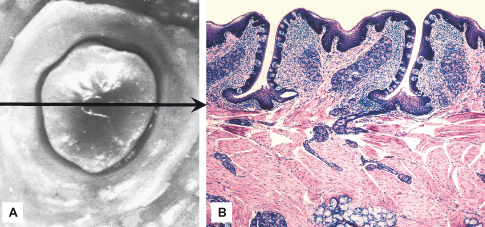
Adjacent and anterior to the sulcus terminalis are 8–12 circumvallate (“walled”) papillae, large structures each surrounded by a deep, circular groove into which open the ducts of minor salivary glands (the glands of von Ebner) (Fig. 6.4). These papillae have a connective tissue core that is covered on the superior surface by a keratinized epithelium. The epithelium covering the lateral walls is non-keratinized and contains numerous taste buds (Fig. 6.4B).
Figure 6.4 The circumvallate papillae (A) are situated in a row anterior to the sulcus terminalis on the dorsal surface of the tongue. (B) A histologic section through the papilla shows the superior surface of the papilla is covered by epithelium with a thin keratinized layer; that of the lateral walls is non-keratinized and contains many taste buds. Ducts of minor salivary glands open between the papillae.
6.2 JUNCTIONS IN THE ORAL MUCOSA
6.2.1 Mucocutaneous Junction
The skin, which contains hair follicles and sebaceous and sweat glands, is continuous with the oral mucosa at the lips (Figs. 6.1 and
Stay updated, free dental videos. Join our Telegram channel

VIDEdental - Online dental courses


