MANDIBULAR THIRD MOLAR
Modern day surgery is based on anatomy. Unless the operator builds on that soild foundation, he is no way better then ‘a hewer of flesh and a drawer of blood’. Hence a discussion of those anatomic structures with which the surgeon is concerned in the surgical removal of mandibular third molar is pertinent.
The mandible consists of a horseshoe shaped body and two flat, broad rami. Each ramus is surmounted by two processes, viz. coronoid process and condylar process.
The lower third molar tooth is situated at the distal end of the body of the mandible where it meets a relatively thin ramus (Fig. 5.1).
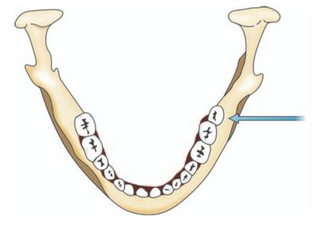
Fig. 5.1: Occlusal view of mandible showing the location of wisdom tooth on left side (blue arrow)
This meeting point constitutes a line of weakness and a fracture may occur if undue force is exerted during elevation of impacted third molar. The tooth is embedded between the thick buccal alveolar bone and a thin lingual cortical plate (Fig.5.2).
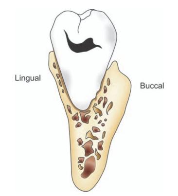
Fig. 5.2: Coronal section of mandible in the region of third molar showing a thick buccal alveolar bone and a thin lingual plate
When the mandible is viewed from below, it will be seen that the wisdom tooth socket lies on a prominent ledge or shelf of lingual bone. In many instances the lingual bone consists of a thin cortical plate less than 1 mm in thickness. Extraction can be facilitated by removal of this thin lingual cortical plate (Fig 5.3). This principle is employed in the lingual split bone technique. In cases where the lingual plate is very thin, attempts to remove fractured apices of tooth may inadvertently lead to their displacement through the lingual plate into the lingual pouch.
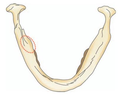
Fig.5.3: View from the inferior surface of mandible to show the lingual shelf of bone (red dotted line) which encloses the mandibular third molar
The buccal bone is predominantly formed by the buccal cortical plate of mandible and the external oblique ridge, the latter being the site of insertion of buccinator muscle. Reduction of the buccal plate will not permit the same ease of surgical access and its loss tends to weaken the mandible. The external oblique ridge is a bulky prominence in some patients and it impedes the buccal surgical approach to the wisdom tooth.
The interdental bone between the second and third molar maybe minimal or even missing. In such case while using elevators extreme care should be used not to damage the bony and periodontal support of second molar, lest it may lead to periodontal pocket formation in the post operative period.
Neurovascular Bundle
Below or alongside the roots of the third molar is the mandibular canal. The canal is usually positioned apically and slightly buccal to the third molar roots. However a variation from the usual position is not infrequent. The canal encloses the neurovascular bundle. The neurovascular bundle contains the inferior alveolar artery, vein and nerve enclosed in a fascial sheath. Since the calcification of the mandibular canal is completed before formation of the roots of third molar, the growing roots may impinge on the canal causing its deflection. Occasionally roots are indented by the mandibular canal, and rarely penetration of the roots of the wisdom tooth by this structure may occur. In the latter case, the neurovascular bundle will be torn during extraction of the tooth. Sometimes the apices may reach the superior wall of the canal and protrude into it (Fig.5.4). In such cases attempted elevation of a small fractured root tip may displace it into the mandibular canal. Furthermore, penetration of mandibular canal by instruments or forceful intrusion of third molar roots may injure the artery or the vein resulting in profuse bleeding.
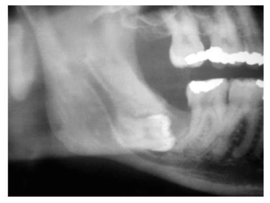
Fig. 5.4: Radiograph showing close relationship of impacted third molar roots to the mandibular canal
From its start at the mandibular foramen, the canal and its contents are surrounded by a thin layer of bone with a configuration similar to lamina dura and this is radiographically detectable. In cases where the roots of the third molar are in direct contact with the neurovascular bundle, the lamina dura may be partially or totally absent. Hence, the radiographic evaluation of the relationship of the mandibular canal and roots of the third molar forms an important part of the preoperative assessment.
Retromolar Triangle
Behind the third molar is a depressed roughened area which is bounded by the lingual and buccal crests of alveolar ridge; the retromolar triangle. Lying lateral to the retromolar triangle is a shallow depression, the retromolar fossa. Either in the retromolar triangle or in the fossa an opening may be present through which emerge branches of the mandibular vessel (Fig.5.5). This branch supplies the temporalis tendon, buccinator muscle and adjacent alveolus. Although these are small vessels, a brisk hemorrhage can occur during the surgical exposure of the third molar region if the distal incision is carried up the ramus and not taken laterally towards the cheek.
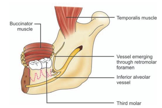
Fig. 5.5: Schematic diagram showing the retromolar vessel emerging through retromolar foramen
The retromolar pad, which is the soft tissue covering the retromolar area is predominantly made up of loose connective tissue. When a gum flap is present over the occlusal surface of third molar, it will resist the upward movement of the tooth during elevation. Therefore a relieving incision through the overlying mucoperiosteum must be made before elevating the tooth.
The tendinous insertion of temporalis muscle terminates as two limiting prongs on the borders of the retromolar triangle. Stripping of these fibers during the removal of third molar can result in postoperative pain.
Facial Artery and Vein
The facial artery and anterior facial vein cross the inferior border of the mandible just anterior to the masseter muscle and have a close relationship to the second and third molar. It is possible to cut these vessels if the BP blade slips when making a buccal cut in that region. Hence, it is always sensible to begin the incision in the depth of the sulcus and direct the blade upwards towards the tooth.
Lingual Nerve
The lingual nerve lies on the medial aspect of the third molar (Fig.5.6)
Fig. 5.6: Schematic diagram showing coronal section through the third molar region and the relationship of important anatomical structures to impacted mandibular third molar
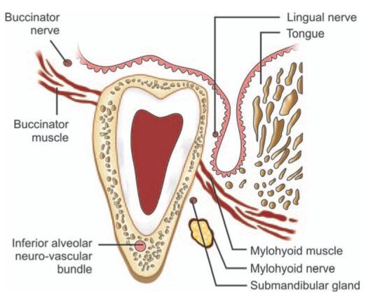
Frequently lingual nerve courses submucosally in contact with the periosteum covering the lingual wall of the third molar socket or it may run below and behind the tooth. The proximity of this important nerve to the third molar places it in danger during the surgical removal of wisdom tooth. Hence great care must be taken to protect it. Injury to lingual nerve will lead to prolonged anesthesia of the anterior two-thirds of the tongue.
Based on studies by Pogrel(1995)1, Holzle(2001)2, Behnia(2000)3 and Keisselbach(1984)4 on cadavers it can be concluded that: (1) the lingual nerve was observed at or above the crest of the lingual plate in 4.6 to 17.6% of the cases; (2) the direct contact of the lingual nerve with the lingual plate in the retromolar region was observed in 22.3 to 62% of the cases; (3) the horizontal distance from the lingual nerve to the lingual plate in these studies ranged from 0 to 7 mm; and (4) vertical distance from the lingual nerve to the crest of the lingual plate ranged from 2 mm above the crest to 14 mm below it.
On an average the lingual nerve is found about 0.6 mm medial to the mandible and about 2.3 mm below the alveolar crest in the frontal plane.
From the above findings it can be concluded that during surgery for the removal of impacted third molar lingual nerve injury is most likely to occur when it traverses along or near the crest of the lingual ridge or in the retromolar pad.
Len Tolstunov (2007)5 cited four anatomical risk factors contributing to lingual nerve injury described below:
1. Lingual version of distoangular impacted lower third molars: This is the first anatomical risk factor to be considered. Distoangular impactions are the most difficult mandibular impacted teeth to extract. The long axis of these third molars is directed away from the operator toward the ramus of the mandible. Radiographically, some of the distoangular impactions show superimposition of roots of the third molar on to the roots of the second molar. Roots of these distoangular impacted mandibular third molars maybe directed lingually. This three-dimensional position of the third molar is often called the lingual version. Thus in distoangular impaction there is vulnerability of the lingual nerve in the retromolar pad area.
2. Lingual plate deficiency: Lingual plate deficiency can present itself as a dehiscence (vertical collapse or cleft) or fenestration below the lingual crest (Fig.5.7). In such cases the apices of third molar penetrates the lingual plate. This indicates the additional risk of deflecting the fractured roots into the lingual pouch during its attempted removal. Its occurrence is likely to be developmental and appears at a time when the third molar forms in the limited space between the vertical ramus of the mandible and the erupted second molar. A pathological lesion in this area (e.g., cyst or tumor) can also erode the lingual plate and further compromise its integrity.
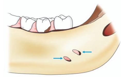
Fig.5.7: Penetration of the apices of roots (blue arrows) of lower third molar through lingual plate
3. High-lateral position of the lingual nerve: High position of the lingual nerve at or above the lingual crest in the retromolar region can place it near or on the lingual plate. Based on studies by various authors quoted above, the lingual nerve can potentially be in full contact with the lingual plate and at or above the crest of the lingual plate up to 2 mm. This again contributes to the vulnerability of the lingual nerve in the retromolar pad area.
4. Local chronic inflammatory condition: Chronic pericoronitis of the lower wisdom teeth is one of the most common reasons for their removal. Occasion/>
Stay updated, free dental videos. Join our Telegram channel

VIDEdental - Online dental courses


