35: Lymphatics and Spread of Dental Infection

Lymphatics and Spread of Dental Infection

LYMPHATIC SYSTEM

The lymphatic system is a part of the overall lymphoid system of the body and a component of the immune system of the body. It is an accumulation of tiny channels or tubules with small nodular structures called lymph nodes interconnecting them. The system functions by returning fluids to the bloodstream from the various tissues of the body. A filtrate of the blood plasma flows out of the capillaries into the surrounding tissues, where it becomes extracellular fluid. It is eventually picked up by the lymphatic vessels or tubules. The extracellular fluid, now referred to as lymphatic fluid, flows through tubules, then through nodes, back through tubules, and possibly through some more nodes. It finally empties into the venous system at the junction of the internal jugular and subclavian veins in the root of the neck and travels back to the heart. This kind of fluid circulation repeats itself continually. The lymph nodes act as filters for the fluids, and the lymphocytes produced within the lymph nodes combat infections that might spread through the lymphatic channels. Most tissues, including the pulp of the teeth, have lymph vessels in them. The distribution of these vessels has been well determined, and this information can be used as a diagnostic tool in the study of oral infections and as a means of slowing the spread of cancers.

Distribution Pattern

We will now examine the distribution pattern of these channels and nodes and discover how they relate to the head and neck area. Fig. 35-1 shows some of the major groups of nodes in the head and neck. The nodes are grouped together into small clusters, which are all interconnected by channels. Each group drains fluids from certain structures or tissue areas, which explains why lymph nodes are involved in combating infections in areas of the body. A sore throat, for example, can be followed by tenderness in the neck and finally a tender lump in that area. In such an instance the infection from the throat has spread through the lymph channels and nodes behind the throat wall known as retropharyngeal nodes. From there it travels through lymph vessels until reaching the first group of lymph nodes in the neck known as upper deep cervical nodes. The lymphocytes in the node have begun to combat the infection and also have started to multiply, causing the node to become enlarged and tender. If the infection is successfully combated in that node, it will subside; however, if the infection is great, it may spread through that lymph node or nodes and on to the next node or group of nodes.

Submandibular nodes

The submandibular nodes are found grouped around the submandibular gland near the angle of the mandible. The easiest way to locate the gland and the nodes is to place a finger on the inferior border of the mandible near the an-gle. Run the finger back and forth until you feel the depression in the inferior border of the mandible. This is the point at which the facial artery and vein cross the inferior border. Just medial to this depression is the submandibular gland, and the submandibular lymph nodes are grouped around it.

The areas that drain into these nodes are all of the maxillary teeth and the maxillary sinus, with the exception of the maxillary third molars; the mandibular canines and all mandibular posterior teeth, with the possibility that the mandibular third molars may not drain here; the floor of the mouth and most of the tongue; the cheek area; the hard palate; and the anterior nasal cavity. As mentioned before, the submental nodes also drain into these nodes. Any infections in these areas tend to cause enlargement and tenderness of the submandibular nodes. This condition is referred to as lymphadenopathy.

Upper deep cervical nodes

The upper deep cervical nodes are located on the lateral surface of the internal jugular vein and lie just beneath the anterior border of the sternomastoid muscle, about 2 inches below the ear. A number of other nodes drain into this group—the submandibular nodes; the nodes behind the back throat wall, known as the retropharyngeal nodes; the parotid nodes in front of the ear and the parotid gland; and others. This is the group affected when you have a particularly sore throat. When a throat infection begins, the first group of nodes involved are the retropharyngeal nodes because they are behind the throat wall. Because it is impractical to palpate these nodes, generally the next group of nodes that are involved in a throat infection, the upper deep cervical nodes, are the first to be noticed. Many people who have sore throats first notice the tenderness in the upper deep cervical nodes. The upper deep cervical group also drains the third molar regions, the base of the tongue, the tonsillar area, the soft palate, and the posterior nasal cavity region. Therefore tenderness in the upper deep cervical nodes without a sore throat should cause you to look in a number of areas to discover the source of the infection.

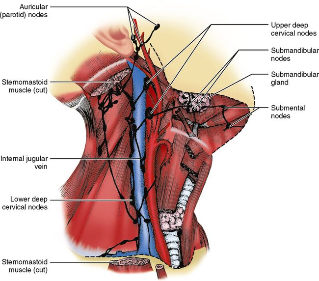
NODE GROUPS AFFECTED BY DISEASE

The terms primary nodes, secondary nodes, and tertiary nodes are often used in discussions about infections and cancer, both of which spread through lymphatic channels. These terms refer to the groups of nodes that are affected in a disease process. If an infection is not stopped by the first (primary) group of nodes, it will spread to the second (secondary) group. If it is not stopped there, it may spread to the third (tertiary) group. One node or group of nodes can be primarily involved in one source of infection, and the same group of nodes can be secondarily or tertiarily involved in another source of infection. Look at the upper deep cervical nodes (see Fig. 35-1). An infection of the third molars may involve these nodes first—the primary group involved. If the infection were in a first molar, the initial sign of infection would be in the submandibular nodes; if it were not successfully combated there, it would spread secondarily to the upper deep cervical nodes. Infections originating in the middle of the lower lip would spread first to the submental nodes, secondarily to the submandibular nodes, and then to the upper deep cervical nodes, which in this instance would be tertiary nodes of involvement. Keep in mind that in infections, any group of nodes may overcome the infection if it is not too severe, and the infection may go no farther.

An understanding of this concept is necessary to comprehend the spread of oral cancer. Each group of nodes acts as a resistance barrier against the spread of cancer. The nodes slow the spread, and if the cancer is detected early enough, it can be treated more successfully. Once the infection or the cancer reaches the lower deep cervical nodes and passes through them, it enters the bloodstream, moving directly into the heart and then throughout the body. With this in mind, it is easy to understand why cancer on the tip of the tongue does not result in as high a mortality rate as does cancer that begins further back on the tongue or in the throat. The tip of the tongue generally drains through four groups of nodes before it enters the bloodstream and spreads throughout the body, whereas cancer in the posterior portion of the tongue or in the throat travels to the upper deep cervical nodes, on to the lower deep cervical nodes, and into the bloodstream. In that area, there are only two groups to stop the spread of the disease. When cancer is detected, knowing the location of the cancer and having a knowledge of the nodes involved allows the surgeon to do a biopsy of the next group of nodes in the chain to see if there is cancer in them. If they are free of cancer, that can be a good sign that the cancer has not spread any further and generally means a more favorable prognosis for the patient.

SPREAD OF INFECTION IN FASCIAL SPACES

Another way through which infections may spread is through fascial spaces. Although infection spread through fascial spaces is much less common, it displays much more dramatic clinical symptoms. The spaces between muscle and tissue layers are referred to as fascial layers or planes, and infections may spread here. You may have seen a patient with a large swollen jaw or with a swollen area beneath the eye. In this situation the infection of dental origin is not spreading through small lymphatic channels but has broken out of the bone around the tooth and is spreading between the tissue or muscle layers.

This kind of infection spread will follow certain predictable pathways, depending on its location. In general, dental infections start in the maxillae or mandible at the apex of a tooth or in the periodontal space around a tooth. Most periodontal space infections cause a swelling of the gingival or mucosal tissue within the oral cavity. Infections at the apices of the teeth cause swelling in one of two directions: buccal or lingual. Most buccal swellings also lead to a swelling in the vestibule of the oral cavity. This swelling is sometimes referred to as a gumboil. The infection comes to a pointed head, breaks through the mucosa, and drains into the oral cavity. If a mandibular infection spreads not in the buccal but in the lingual direction, it will travel to the tissue spaces in two specific areas—above the mylohyoid muscle in the floor of the mouth, or beneath the mylohyoid muscle in the tissue beneath the chin—depending on its point of origin.

How can one predict where the infection will go? Refer to Fig. 26-17, a medial view of the body of the mandible, and picture the lengths of the roots of the individual teeth. Look at the mylohyoid line on the mandible and notice its location relative to the apices of the roots. You can see that, in general, the apices of the mandibular molar teeth are inferior to the mylohyoid line, whereas the apices of the roots of premolars and the anterior teeth are above the mylohyoid line. Therefore a molar infection will tend to break out of the bone below the mylohyoid line and spread to the space beneath the chin, referred to as the submental space. Infections of the premolars and the anterior teeth will tend to break out of bone above the mylohyoid line and spread to the spaces in the floor of the mouth, referred to as the sublingual space.

Infection spreading into the sublingual space causes a swelling into the floor of the mouth. If it spreads into the submental space, it will cause a swelling beneath the chin, sometimes referred to as Ludwig’s angina. These infections continue to spread by gravity if not treated. Whether above or below the mylohyoid muscle, as they spread down and back, they reach the posterior end of the mylohyoid muscle. Both kinds of infection eventually reach the side of the neck next to the pharynx, which is referred to as the lateral pharyngeal or parapharyngeal space. This causes swelling on the side of the neck if left untreated. From here the infection may spread around the pharynx to its posterior border, which is referred to as the retropharyngeal space, and from there to the posterior mediastinum, which is in the back of the chest or thoracic cavity. If the infection reaches this point, the person may die within a short period of time. With the advent of antibiotics, these infections are not so frequently seen as they were in the past but can occasionally still be found.

The importance of this section is not to be able to completely describe or define the boundaries of these spaces or potential spaces, but to understand how the origin or location of the original infection determines the pathway it will follow and the potential outcome if left untreated.

OTHER MAXILLARY INFECTIONS

Maxillary infections react slightly differently because of the anatomic features of the area. If the infection does not open into the maxillary buccal vestibule or onto the palate, it may spread toward three areas—the nasal cavity, the maxillary sinus, or the soft-tissue spaces of the cheek or the area below the eye. The area involved is related to the tooth involved. A swelling below the eye is usually related to infection from an anterior tooth, usually the maxillary canine, whereas swelling in the cheek is usually related to infection in a posterior tooth. Although it is possible for infection to spread to the nasal cavity or maxillary sinus, it is rather rare. These maxillary infections around the eye or cheek can also spread to the lateral pharyngeal space and from there to other areas.

Stay updated, free dental videos. Join our Telegram channel

Jan 4, 2015 | Posted by in General Dentistry | Comments Off on 35: Lymphatics and Spread of Dental Infection

VIDEdental - Online dental courses

Get VIDEdental app for watching clinical videos