Tooth Identification
Following is a description of general characteristics of each of the teeth by their respective groupings: incisors, canines, premolars, and molars.* In identifying teeth, it is necessary to be able to differentiate among the left and right teeth in any particular group.
GENERAL RULES OF TOOTH IDENTIFICATION
1. The curvature of the cementoenamel junction (CEJ) is usually about 1 mm less on the distal surface of the tooth than on the mesial.
2. Tooth roots do not always curve; however, if they do curve, they usually curve distally, especially at the apex of the root. It is not uncommon, however, for the root to curve toward mesially.
3. The distal incisal edges of anterior teeth are more rounded than the mesial incisal edges.
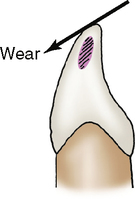
4. Mandibular anterior teeth tend to wear on their labial incisal edges, whereas maxillary teeth wear on their lingual incisal edges. Unless a person has a class III occlusion, the maxillary teeth are facial to the mandibular teeth.
5. Permanent molars are generally smaller in height and have fewer cusps the more posteriorly they are positioned. For example, the permanent first molar usually has five cusps and is larger than a second or third molar. A mandibular first molar has a distal cusp on its facial surface, and a maxillary first molar has a cusp of Carabelli. The second and third molars are less likely to have these cusps; however, when they do, cusps are less well developed and are more like tubercles than cusps.
6. Permanent molars tend to have more secondary and tertiary anatomy the more posterior they are positioned. Secondary anatomy consists of extra grooves and pits in addition to the main primary developmental anatomy. These grooves and pits are more shallow than the primary anatomy and are more likely to be found on second and third molars. Tertiary anatomy refers to the extremely shallow and even more numerous grooves, pits and lines that third molars often have, giving them a more wrinkled appearance than first or second molars.
7. The roots of molars tend to be shorter and closer together the more posterior the molars are positioned, and the roots are often fused into one. First molars have the widest and longest roots of all molars.
8. The more posterior the molars are positioned, then the more variation of the anatomy is evident. Third molars are more wrinkled and unpredictable in shape than second or first molars; they could have six cusps or one single conical cusp. They are also more likely to be congenitally missing than other molars.
INCISORS
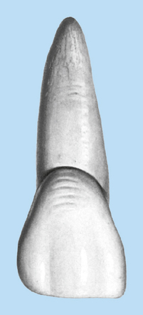
1. Incisal two thirds appear flattened on labial and lingual sides (Fig. 10-1)
Maxillary
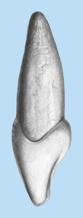
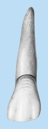
1. Crown wider mesiodistally than faciolingually (Figs. 10-3 and 10-4)
2. Root has triangular cross section, being broader on facial side
Central
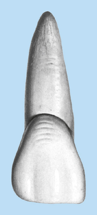
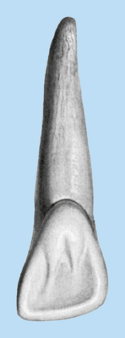
1. Greater crown-to-root ratio than lateral incisors (crown larger, root about same as or smaller than lateral incisors) (Figs. 10-5 and 10-6)
2. Mesioincisal angle relatively sharp (90-degree angle), with contact area in incisal third
Lateral
1. Lesser crown-to-root ratio than central incisors (crown smaller, root about same as or larger than central incisors)
2. Mesioincisal angle rounded, with contact area at junction of middle and incisal thirds
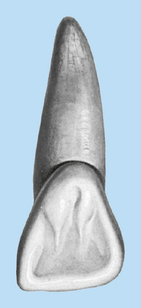
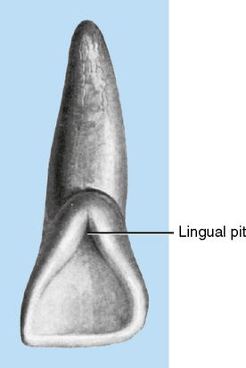
3. Small cingulum, often with a lingual pit (Figs. 10-7 and 10-8)
Right-left
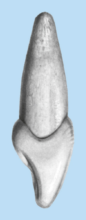
1. Mesioincisal angles more square than distoincisal angles (Fig. 10-9)
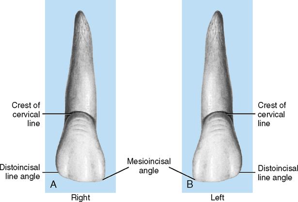
2. Crest of cervical line more often displaced toward distal from labial or lingual view
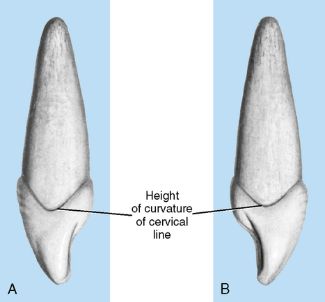
3. Mesiocervical line curves more incisally than distocervical line (Figs. 10-10)
Mandibular
1. Smaller than maxillary central or lateral incisor (Figs. 10-11 and 10-12)
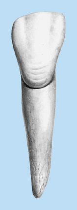
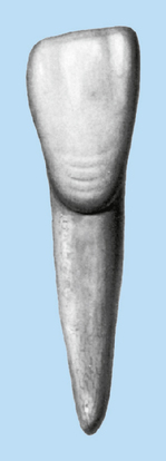
2. Crown wider faciolingually than mesiodistally
3. Root with oval cross section
4. Incisal edge wears on labial surface (Fig. 10-13); incisal edge angled toward the lingual side
Stay updated, free dental videos. Join our Telegram channel
VIDEdental - Online dental courses


