Fundamental and Preventive Curvatures
Proximal Alignment of the Teeth and Protection of the Periodontium
EVOLUTION OF FUNDAMENTAL AND PREVENTIVE CURVATURES AND PROXIMAL ALIGNMENT OF THE TEETH
1. Specific location and size of proximal (mesial or distal) contact areas of various teeth
2. Size and location of interproximal spaces formed by the proximal contact surfaces
3. Location and effectiveness of the embrasures or spillways
4. Facial and lingual contours on the labial, buccal, and lingual surfaces of crowns
5. Amount of curvature of the cementoenamel junction (CEJ) on the mesial and distal surfaces of the various teeth
6. Self-cleaning qualities of the tooth, smoothness of the enamel, and overall shape of the tooth to meet its function
Proximal Contact Areas
Fig. 3-1 shows the contact areas of two premolars. The contact area on the distal surface of the first premolar is called the distal contact area. What would the contact area on the mesial surface of the second premolar be called? The contact area is not just a point but rather a flattened portion of the tooth where it actually touches the tooth next to it in the same dental arch.
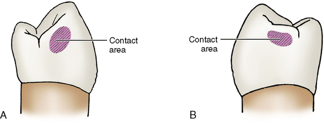
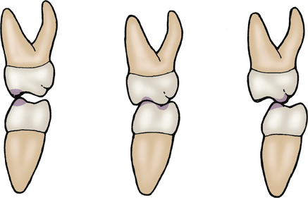
A contact point differs from a contact area. A contact point is where the occlusal cusp of one tooth touches the occlusal portion of another tooth in the opposing arch (Fig. 3-2). The contact point of an upper tooth hits (occludes and makes contact with) the contact point of a lower tooth.
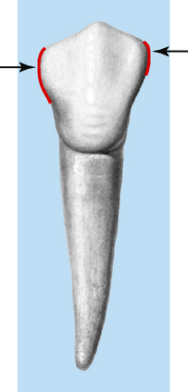
Looking at a buccal view of a tooth (Fig. 3-3), notice that the contact area occurs at the portion of the tooth that has the greatest curvature. In other words, the distal contact area occurs at the part of the distal portion of the tooth that bulges or curves out the most.
Interproximal Spaces
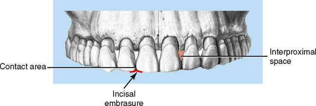
Interproximal spaces are triangular-shaped spaces between the teeth formed by the bone on one side and the proximal surfaces and their contact area on the other side (Fig. 3-4). The contact area forms the apex of the triangle, the proximal surfaces form the sides, and the alveolar bone makes up the base. These spaces are normally filled with gingival tissue called papillary gingiva or interdental papilla. By its presence the interdental papilla keeps food from collecting cervically to the contact areas between the teeth. The interdental space (space between teeth) provides a place for a bulk of bone, thus affording better anchorage and support. This space is wider cervically than occlusally to provide more access for the vascular support to nourish the interdental bone and papillary tissue. This also affords a stronger bony base. When gingival recession occurs between the teeth, the interdental papilla and bone no longer fill the entire interproximal space; a void exists cervically to the contact area. This void is called a cervical embrasure. The more bone that is missing, then the larger is the interproximal space because bone forms the base of the latter. The cervical embrasure could become larger because gum tissue receded while the bone level stayed the same. In this case the interproximal space would remain the same. Cervical embrasures occur often as a pathologic consequence of periodontal or orthodontic causes, and these embrasures offer a place in which bacteria, calculus, and food debris can accumulate (see Fig. 3-4).
Embrasures
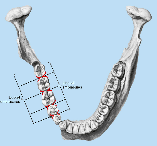
Embrasures (spillways) are the spaces between the teeth that are occlusal to the contact areas (Fig. 3-5). They allow for the passage of food around the teeth so that food is not forced into the contact area between the teeth. These embrasures are named for their location in relation to the contact area. For instance, the space buccal to the contact area is the buccal embrasure; the lingual embrasure is lingual to the contact area. The names of the embrasures are facial (buccal or labial), lingual, incisal, or occlusal. Gingival embrasures are also evident, but only if the interproximal space is not occupied by any gingiva or bone. The gingival embrasure is gingival to the contact area and not usually present; it is the same as the cervical embrasure discussed in the last section. The embrasures have the following purposes:
Stay updated, free dental videos. Join our Telegram channel
VIDEdental - Online dental courses


