The Tongue
• To describe the formation of the tongue as it relates to the germ layers and its pharyngeal arches of origin
• To discuss the difference between extrinsic and intrinsic muscles of the tongue
• To describe briefly how tongue movement is accomplished
• To describe the papillae of the tongue and their function
• To describe the kinds of changes seen on the tongue that indicate health problems
DEVELOPMENT OF THE TONGUE
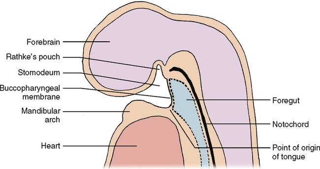
As discussed in Chapter 18, a number of bars of tissue are found on the anterior surface of the developing embryo, which are referred to as pharyngeal arches. The first pharyngeal arch is the mandibular arch, the second is the hyoid arch, and the remainder are numbered III, IV, and VI (V disappears). Just above the first arch and extending down behind all the arches is a hollow tube, the digestive tract. Before the end of the fourth embryonic week, this tube is closed off at the upper end by the buccopharyngeal membrane, which separates the upper end or foregut from the primitive oral cavity or stomodeum. The epithelium anterior to the buccopharyngeal membrane develops from the outer germ layer or ectoderm. The tube behind the buccopharyngeal membrane develops from the inner germ layer or entoderm. At about 4½ weeks the buccopharyngeal membrane ruptures, but the epithelium in that area still comes from two distinct germ layers. It is at this point that the tongue starts to develop as a swelling that arises out of the back part of the pharyngeal arches (Fig. 24-1). This swelling develops from the future floor of the mouth. The epithelial covering of the tongue develops from ectoderm and entoderm—the anterior two thirds from ectoderm and the posterior one third from entoderm. The tongue is basically a sac of epithelium filled with muscles. These muscles arise from the middle germ layer of the embryo, the mesoderm.
Each pharyngeal arch is associated with a particular cranial nerve; Chapter 34 on the nervous system discusses from which pharyngeal arch the various parts of the tongue develop. If you refer back to Chapter 18 you will find that the anterior two thirds of the tongue develops from two lateral lingual swellings and a midline tuberculum impar. These are both from the first arch. The posterior one third of the tongue develops from the copula (hypobranchial eminence) and the third arch. The root of the tongue and epiglottis develops from the epiglottal swelling of the fourth arch.
TONGUE MUSCLES
1. Superior longitudinal group—runs from front to back (anterior to posterior) and lies near the dorsum of the tongue
2. Inferior longitudinal group—also runs anterior to posterior but lies near the bottom or ventrum of the tongue
3. Transverse group—runs from side to side
4. Vertical group—runs from top to bottom (dorsal to ventral)
Stay updated, free dental videos. Join our Telegram channel
VIDEdental - Online dental courses


