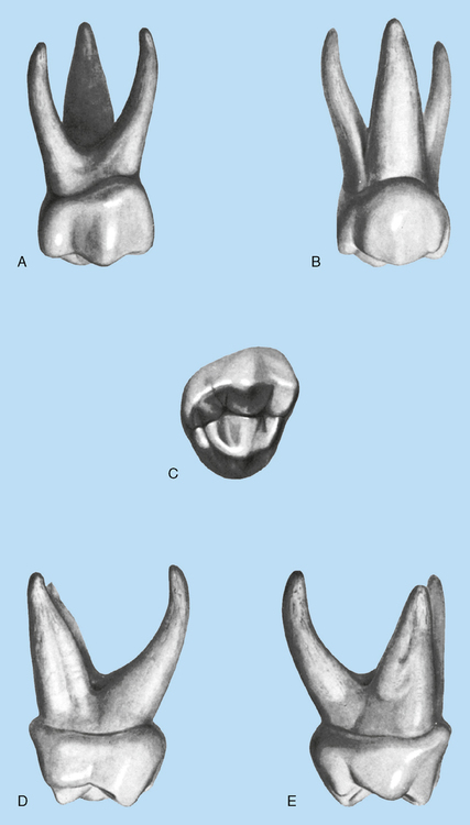
Deciduous Dentition
• To identify the various deciduous teeth
• To recognize whether a tooth is primary or secondary
• To know the eruption dates of the primary and secondary teeth
• To understand the essential differences between deciduous and permanent teeth
• To understand the importance and functions of deciduous teeth
• To compare the dental anatomic features of deciduous teeth, not only with the other deciduous teeth but also with their permanent counterparts
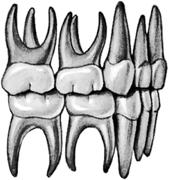
The primary or deciduous dentition consists of 20 teeth, each quadrant containing two incisors, one canine, and two molars (Fig. 16-1).
The first deciduous teeth to erupt, about 8 months after birth, are the mandibular central incisors. The maxillary central incisors usually erupt about a month later. As in the permanent teeth, the primary mandibular teeth usually erupt before the maxillary. The following is an approximate eruption schedule of the deciduous teeth (see Fig. 5-7).
Central incisors: 6 to 12 months
ESSENTIAL DIFFERENCES BETWEEN DECIDUOUS AND PERMANENT TEETH
1. The deciduous anteriors are smaller than their permanent successors in both their crown and root proportions. Deciduous molars are wider mesiodistally than the permanent premolars, which will take their places.
2. The roots of deciduous anterior teeth appear longer and more slender proportionately when compared to permanent teeth. All permanent teeth have much longer roots but the crowns of deciduous teeth are so short that proportionately their roots appear to be long and slender.
3. The roots of deciduous posterior teeth are very narrow at their cementoenamel junctions (CEJs) where the crowns join the roots. In addition the root trunks of deciduous molars are very short.
4. The cervical ridge of enamel at the cervical third of the anterior crown labially and lingually is much more prominent in deciduous dentition. These bulky ridges extend out from the very narrow cervical necks of the teeth.
5. The buccocervical ridges on the deciduous molars are much more pronounced, especially on first molars. These cervical prominences give deciduous crowns a bulbous appearance and accentuate the narrow cervical portion of deciduous roots.
6. The buccal and lingual surfaces of deciduous molars taper occlusally above the cervical curvatures much more so than the permanent molar buccal and lingual surfaces. This results in a much narrower occlusal table of the occlusal surface buccolingually.
7. The roots of the deciduous molars when compared to permanent molars are also proportionately longer and more narrow because the deciduous crowns are so short. These roots also flare apically to allow room for the permanent teeth to develop between them (Fig. 16.2).
8. The deciduous teeth are usually lighter in color than the permanent teeth. They have a whiter color with a bluish cast. Permanent teeth have more yellow, grey, or brown tones.
9. The pulp chambers of deciduous teeth are relatively large in comparison with the crowns that envelop them.
10. The pulp horns of deciduous teeth extend rather high occlusally, placing them much closer to the enamel than the pulp horns in permanent teeth.
11. The dentin thickness between the pulp chambers and the enamel is much thinner than in permanent teeth.
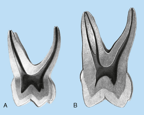
12. The enamel of deciduous teeth is relatively thin and has a consistent depth (Fig. 16-3).
THE IMPORTANCE OF DECIDUOUS TEETH
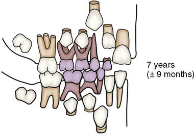
The spaces between the deciduous canines and first molars and those between the first and second molars are called leeway spaces. They allow an extra margin of space for the eruption of the permanent canine, and the first and second premolars. Leeway space is necessary because several offsetting factors are present. First, the mesiodistal measurement of the two permanent premolars combined is less than the sum of the mesiodistal measurements of the deciduous molars. Although this allows extra room for the premolars, the permanent canine requires more room than the deciduous canine. Second, bone growth allows for leeway space, but this is offset by the phenomenon of mesial drift. The first permanent molar tends to move mesially; thus the amount of space reserved for the permanent premolars is shortened. If a deciduous molar is prematurely lost or a decayed interproximal space is not restored, a permanent molar pushes into this space and blocks out the premolar. Little, if any, extra space is available (see Fig. 16-3).
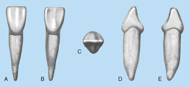
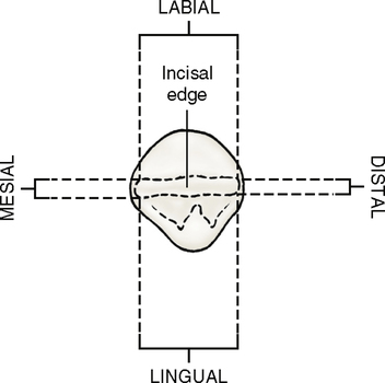
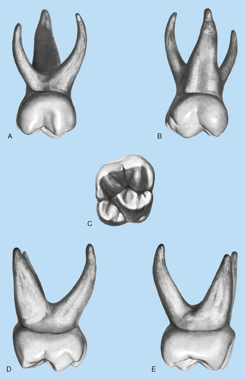
Maxillary Central Incisors (Fig. 16-4)
Labial aspect (Fig. 16-5; see Fig. 16-4, A)
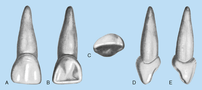
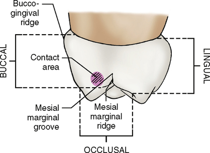
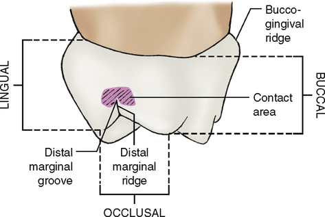
Mesial and distal aspects
From the proximal aspects (Fig. 16-7; see Fig. 16-4, D and E) the crown appears wide in relation to its total length. Because of its short length, the labiolingual measurements make the crown appear thick, even at the incisal third. The mesiocervical curvature is greater than the distal curvature.
Maxillary Lateral Incisors
A lateral incisor’s crown is smaller than a central incisor’s crown in all dimensions, except that the cervicoincisal length is greater than its mesiodistal width. In all other ways, it appears similar to a central incisor. The root appears much longer in proportion to the crown when compared with the central (Fig. 16-8).
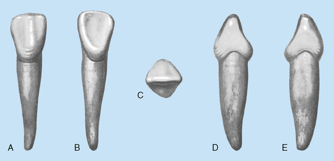
Mandibular Central Incisors (Fig. 16-9)
Labial aspect
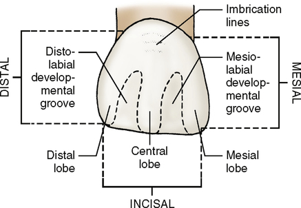
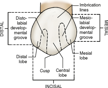
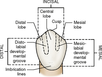
Mamelons or grooves may be visible in a labial view of a deciduous mandibular incisor (Fig. 16-10). The crown appears wide in comparison with its permanent successor. The mesial and distal sides of the crown taper evenly from the contact areas. The root may be two to three times the height of the crown. It is very narrow and is also conical in shape.
Mesial and distal aspects
From the mesial aspect the incisal ridge is centered over the root. The labial and lingual cervical contours are quite convex, much more so than those of the permanent mandibular incisors. Cervical curvature is greater on the mesial side than on the distal side (Fig. 16-12; see Fig. 16-9, D and E).
Mandibular Lateral Incisors
The mandibular lateral incisors (Fig. 16-14) are wider and longer than the central incisors, and their cingula are more developed. Labiolingually the lateral incisors are also wider. There is a tendency for the incisal ridge to slope distally, and its distal margin is more rounded.
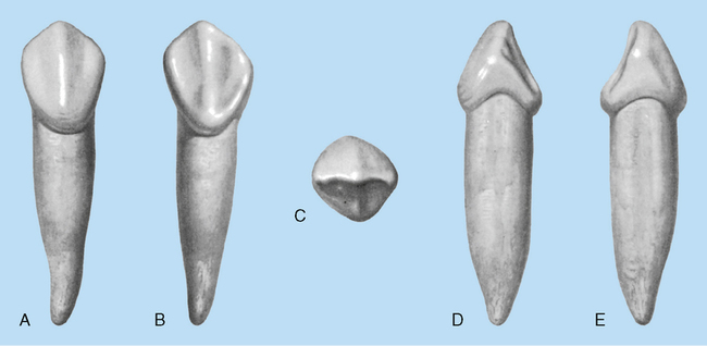
Maxillary Canines (Fig. 16-15)
Labial aspect (Fig. 16-16; see Fig. 16-15, A)
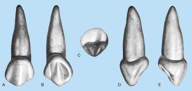
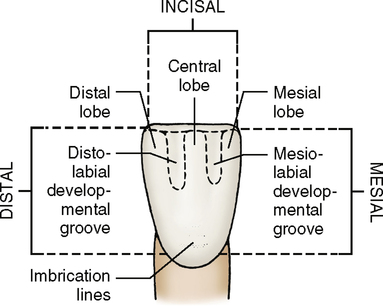
Lingual aspect
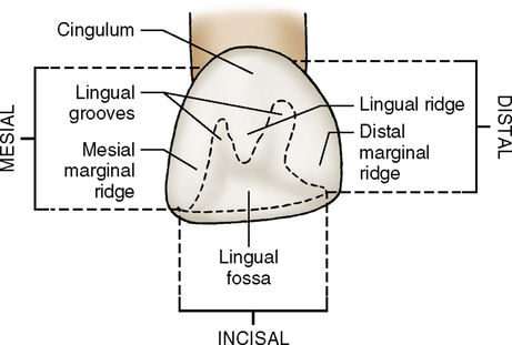
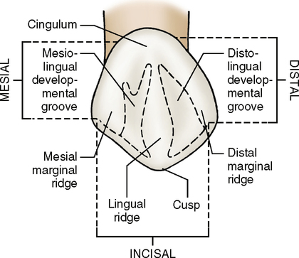
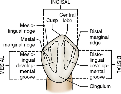
From the lingual view (Fig. 16-17; see Fig. 16-15, B) the mesial and distal marginal ridges, incisal ridges, and cingulum are all very pronounced. A tubercle may extend from the cusp tip to the lingual ridge. The lingual ridge extends from the cusp tip to the cingulum and divides the lingual surface into mesiolingual and distolingual fossae.
Mandibular Canines (Fig. 16-20)
Labial aspect
Compared with a maxillary canine, the labial surface of a mandibular canine (Fig. 16-21; see Fig. 16-20, A) is much flatter, with shallow developmental grooves. The distal cusp ridge is longer than that of a maxillary canine. The root is long, narrow, and almost twice the length of the crown, although it is shorter and more tapered than that of a maxillary canine.
Lingual aspect
The most obvious difference between the maxillary and mandibular canines is the presence of a slight concavity called the lingual fossa. Instead of two lingual fossae, one is present. The lingual surface (Fig. 16-22; see Fig. 16-20, B) is less prominent than that of a maxillary canine, and the crown converges lingually so that it is narrower on the lingual side than on the labial.
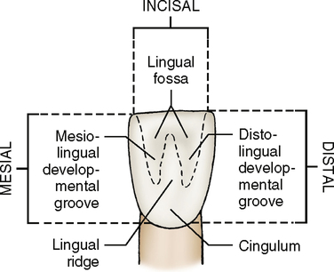
Incisal aspect
The incisal ridge (Fig. 16-24; see Fig. 16-20, C) is straight and centers over the crown labiolingually. The lingual surface shows a definite tapering toward the cingulum. The labial surface from this aspect presents a flat surface with a slight convexity, whereas the lingual surface presents a flattened surface that is slightly concave.
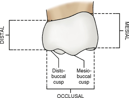
Maxillary First Molars (Fig. 16-25)
Buccal aspect (Fig. 16-26; see Fig. 16-25, A)
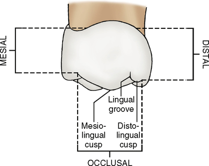
Lingual aspect
The lingual view (Fig. 16-27; see Fig. 16-25, B) shows that the crown of a deciduous maxillary first molar converges toward the lingual surface.
Occlusal aspect
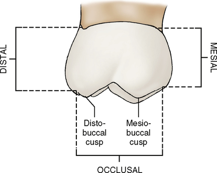
The occlusal view (Fig. 16-30; see Fig. 16-25, C) shows that the crown converges in a lingual direction so that the occlusal table appears triangular. The crown may have three or four cusps. If four are present, then two are on the buccal side and two are on the lingual. If three cusps develop, only one is lingual.

Maxillary Second Molars (Fig. 16-31)
Buccal aspect
A deciduous maxillary second molar resembles a permanent maxillary first molar, although it is much smaller. From the buccal view (Fig. 16-32; see Fig. 16-31, A) two equal-sized buccal cusps with a buccal groove between them are visible. As on a deciduous first molar, the crown is narrow at its cervix, compared with its mesiodistal measurement at the contact area. A deciduous second molar is much larger than a deciduous first molar both in crown and root formation. The two buccal cusps are about equal in size. How is this different from the cusps of a deciduous first molar?
Stay updated, free dental videos. Join our Telegram channel
VIDEdental - Online dental courses


