Nose, Nasal Cavity, and Paranasal Sinuses
• To define the terms nose, nasal cavity, and paranasal sinuses
• To understand the anatomy of the nose, nasal cavity, and paranasal sinuses
• To describe the function of the nasal cavity, nasal epithelium, and paranasal sinuses
• To discuss the anatomic relationship of the maxillary sinus and the maxillary teeth
• To describe the relationship of maxillary teeth to the maxillary sinus in infections of either one
NOSE AND NASAL CAVITY
External View
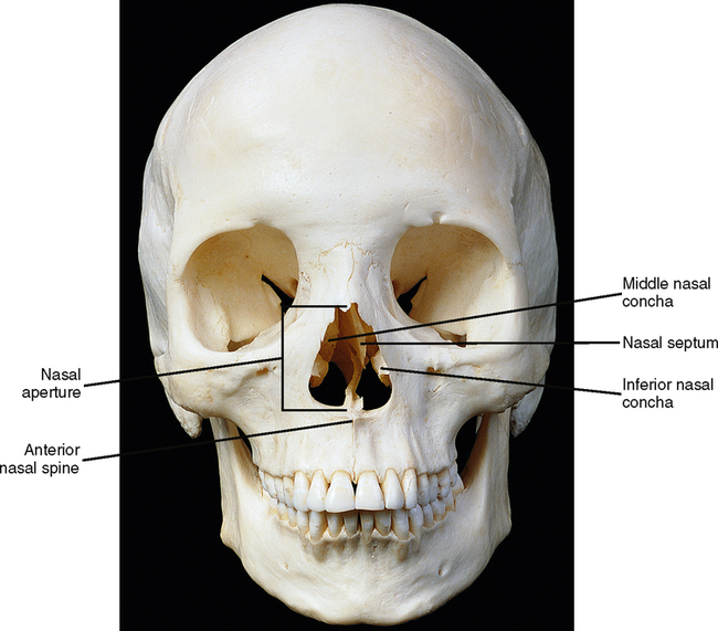
The nose and nasal cavity are a complex arrangement of hard and soft tissues. The nose is that portion of the nasal complex that protrudes outward from the skeletal component. The nose, or more properly, the external nose, is attached superiorly to the nasal bones and inferiorly to the anterior nasal spine. Each protruding lateral margin is known individually as a wing of the nose, or an ala. The external nose is divided in half by the cartilaginous part of the nasal septum, which is the wall that divides the nasal complex into right and left halves (Fig. 27-1). If you look at a skull, you will see that the nasal aperture, the anterior most portion of the nasal cavity, is somewhat pear shaped.
Internal View
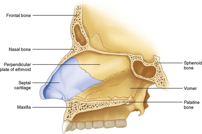
The septum is formed from the vomer bone and a portion of the ethmoid bone as well as the fibrocartilaginous part of the septum (Fig. 27-2). The inferior portion or floor of the cavity is formed from the bones of the hard palate, which are the palatal processes of the maxillae and the horizontal portion of the palatine bones. The most complicated portion of the nasal cavity is its lateral walls. The upper half to two thirds of the lateral wall of the cavity is formed from parts of the ethmoid bone. It consists of the wall and two shelflike structures known as the superior and middle nasal conchae. The lower part of the lateral walls are formed by portions of the maxillae. At the point where the maxillae and the ethmoid bone meet in the lateral wall, there is a third medial projection, which is a separate bone itself, known as the inferior nasal concha (Fig. 27-3).
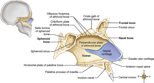
As you reach the posterior part of the nasal septum, you come to an area known as the choana, or posterior nasal aperture. Posterior to this is the nasal pharynx, which is discussed in Chapter 31. The posterior-superior part of the nasal cavity is composed of a portion of the body of the sphenoid bone where it meets the ethmoid bone and is known as the sphenoethmoidal recess. Visualizing this from a sagittal view of a skull, the sphenoidal sinus is below the pituitary fossa.
Epithelial Lining
Recall from Chapter 17 on epithelial tissues that there is one type of epithelium known as pseudostratified columnar epithelium with goblet cells. This epithelial tissue is usually called respiratory epithelium because it is primarily found in the respiratory tract. (You should be aware by now that the nasal cavity is the beginning of the respiratory tract or system.) The epithelium has many hairlike projections known as cilia that move in a synchronized beating pattern toward the anterior portion of the nasal cavity. The tiny goblet cells secrete a sticky mucous substance onto the cilia, trapping contaminants as they enter the nasal cavity and moving them toward the front where they are removed by blowing the nose; during sleep, this mucus may flow backward as postnasal drip. For this mechanism to be successful it is important to have as much surface area as possible in the nasal cavity. The conchae help accomplish that.
Stay updated, free dental videos. Join our Telegram channel
VIDEdental - Online dental courses


