3
Growth and development of the neurocranium
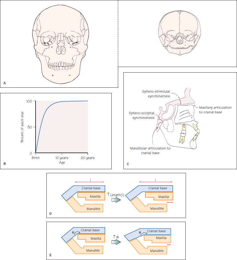
Figure 3.1 (A) In comparison to an adult skull, that of a new born child has a much larger cranial vault in proportion to the facial skeleton because of the relatively advanced neural growth. (B) A growth curve for the cranial vault. (C) The location of the main cranial base synchondroses and the relationship of these to the articulation of the maxilla and mandible. (D) The influence of cranial base length on the relationship between the maxilla and mandible. With an increase in cranial base length, there is a tendency towards a skeletal II pattern. When the length reduces, the skeletal pattern is likely to tend towards Class III. (E) The influence of cranial base angle on the skeletal relationship. With an increase in cranial base angle, there is a tendency towards a skeletal II pattern. When the angle reduces, the skeletal pattern is likely to tend towards a Class III relationship.

Cranial vault
The cranial vault is formed by the frontal, parietal, squamous part of the temporal and occipital bones. All these bones form by intramembranous ossification of the mesenchymal tissue surrounding the developing brain, with centres of ossification first appearing at approximately 8 weeks in utero. Sutures, which are periosteum-lined fibrous joints, separate the individual bones and provide tension-adapted growth. Fontanelles are found where sutures merge and increase the flexibility of the cranial vault to facilitate its passage through the birth canal during parturition. All the fontanelles are closed by the age of 18 months.
Growth of the cranial vault is largely determined by growth of the underlying brain. As brain growth is advanced at an/>
Stay updated, free dental videos. Join our Telegram channel

VIDEdental - Online dental courses


