14
Extra-oral examination: skeletal pattern
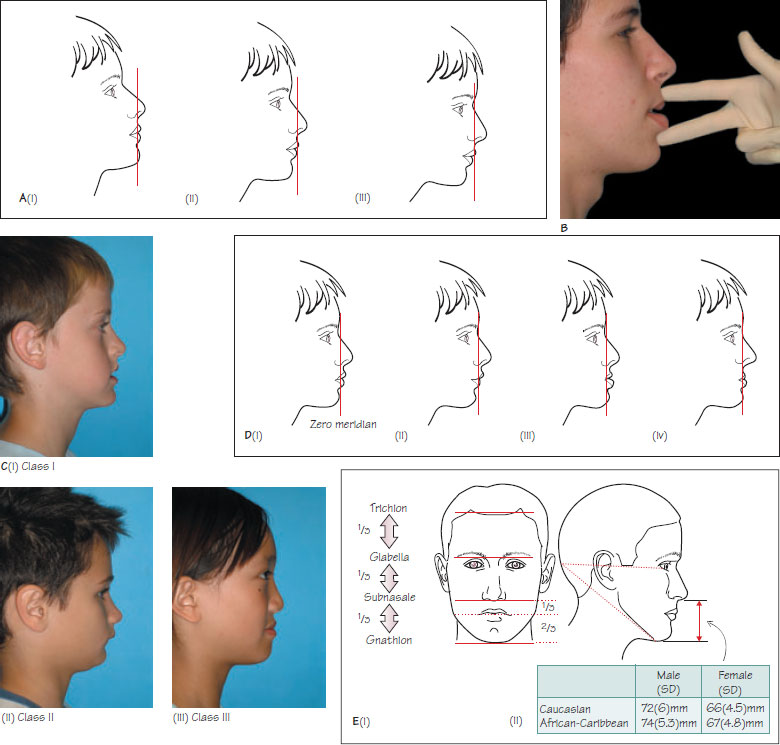
Figure 14.1 (A) The orientation of the head can affect the assessment of the AP skeletal pattern. Ideally, the head should be in the natural head position (ii) to achieve this, the patient should be sitting upright, relaxed, and looking straight ahead at a distant point at eye level. Incorrect head posture can lead to an inaccurate assessment of skeletal pattern (i, iii). (B) Assessment of the AP skeletal pattern. (C) An example of a (i) skeletal I, (ii) skeletal II and (iii) skeletal III pattern. (D) The zero meridian can be used to assess AP jaw position in relationship to the cranial base and gives an indication of which jaw(s) is contributing to the skeletal discrepancy. (i) Normally, the base of the upper lip should lie 2–3 mm ahead and the lower lip 0–2 mm behind this line. (ii) Class II pattern due to mandibular retrognathia. (iii) Class III pattern due to mandibular protrusion. (iv) Class III pattern due to maxillary retrusion. (E) Assessment of the vertical dimension by (i) examining the proportional relationship of the LAFH to the mid-face and (ii) assessment of the FMPA and absolute LAFH.
Orthodontic examination should begin as soon as the patient enters the surgery. The general stage of development, of which statural height is a good indicator, should be noted. The presence of secondary sexual characteristics (Figure 2.1D) also provides a good indication of developmental stage. This chapter will focus on the assessment of skeletal pattern.
Assessment of skeletal pattern
The relative position of the maxilla and mandible, termed the skeletal pattern, has a large influence on the relationship of the maxillary and mandibular dentitions. The skeletal pattern should be assessed in three dimensions:
- Anteroposterior (AP);
- Vertical;
- Transverse.
Anteroposterior dimension
The aim is to relate the AP position of the mandible to the maxilla and the relationship of these bones to the cranial base. Assessment of the position of each jaw relative to the cranial base gives an indication of which jaw has contributed to any discrepancy. It is important to assess the patient in the natural head position, which is a standardised reproducible head orientation, as the tilt of the head can influence the interpretation of skeletal pattern (Figure 14.1A). To achieve this, the patient should be sitting/>
Stay updated, free dental videos. Join our Telegram channel
VIDEdental - Online dental courses


