27
The nasal cavity and paranasal sinuses
Chapter contents
27.1 Introduction
The nasal cavity is the entrance to the respiratory tract. Its functions are to clean, warm, and humidify air as it is inhaled. Respiratory mucosa covered by pseudostratified ciliated epithelium and goblet cells, as described in Chapter 5 and illustrated in Figure 5.2B, lines the majority of the nasal cavity. The cilia and mucus trap particles, thus cleaning the air; the mucus also humidifies the air and warming is achieved through heat exchange from blood in the very vascular mucosa. The efficiency of all these processes is increased by expanding the surface of the nasal cavity by folds of bone. The nasal cavity also houses the olfactory mucosa for the special sense of olfaction although the olfactory mucosa occupies a very small proportion of the surface of the nasal cavity.
The nasal cavity extends from the nostrils on the lower aspect of the external nose to the two posterior nasal apertures between the medial pterygoid plates where it is in continuation with the nasopharynx. Bear in mind that in dried or model skulls, the nasal cavity is smaller from front to back and the anterior nasal apertures seem extremely large because the cartilaginous skeleton of the external nose is lost during preparation of dried skulls.
As you can see in Figure 27.1, the nasal cavity extends vertically from the cribriform plate of the ethmoid at about the level of the orbital roof above to the palate, separating it from the oral cavity below. Figure 27.1 also shows that the nasal cavity is relatively narrow from side to side, especially in its upper part between the two orbits and widens where it sits between the right and left sides of the upper jaw below the orbits. The nasal cavity is completely divided into right and left compartments by the nasal septum. From the anterior view seen in Figure 27.1, you can see that the surface area of lateral walls of the nasal cavity are extended by the three folds of bone, the nasal conchae.
27.2 The external nose
The skeleton of the external nose shown in Figure 27.2 comprises the nasal bones, the upper and lower nasal cartilages, the septal cartilage, and the cartilaginous part of the nasal septum. The nasal bones and adjacent parts of the frontal processes of the maxillae form the bridge of the nose. The upper and lower nasal cartilages with a variable number of minor cartilages form the skeleton of the lower pliable part of the external nose. As seen in Figure 27.1B, the septal cartilage is quadrilateral and is continuous posteriorly with the anterior edge of the perpendicular plate of the ethmoid and the superior border of the vomer, the bones that form the bony nasal septum (see p. 287). The various cartilages are unmineralized parts of the nasal capsule from which they develop (see Section 33.3).
The lateral border of each external nostril is thickened and rounded by the presence of fatty fibrous tissue. The nasal cavity is dilated just inside the external nostrils to form the nasal vestibule which is lined by hairy skin with coarse hairs that filter large particles.
The sensory innervation of the external nose skin is by cutaneous branches of the ophthalmic and maxillary trigeminal nerves and its arterial supply is through branches of the facial, ophthalmic, and infraorbital arteries. The venous drainage is into corresponding veins and the lymphatic drainage is into the submandibular nodes.
27.3 The nasal cavity
The skeletal structure of the nasal cavity is illustrated in Figure 27.3. You will need to examine a skull sectioned in the sagittal plane to see clearly the internal structure of the nasal cavity and the bones that form it, but you may be able to see some features on an intact skull. Be aware that many of the bones forming the nasal walls are quite delicate and may be broken off dried skulls.
Figure 27.3 illustrates that the roof of the nasal cavity is formed by the nasal bones anteriorly, the cribriform plate of the ethmoid in its intermediate part, and by the underside of the body of the sphenoid posteriorly. Its floor is the bony palate made up of the palatine processes of the maxillae in front and horizontal plates of the palatine bones behind. Each palatine process is pierced anteriorly, close to the septum, by an incisive canal which transmits the nasopalatine nerves and the terminal branch of the greater palatine artery. The two canals open into the incisive fossa, a single funnel-shaped depression on the oral surface of the palate (see Figure 25.4).
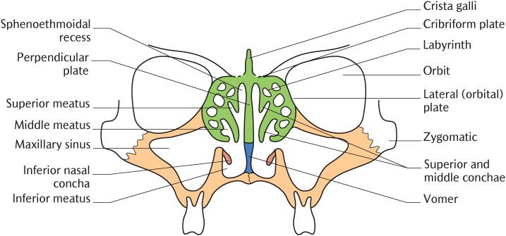
Fig. 27.1 Diagrammatic representation of the contributions of the ethmoid and maxilla bone to the lateral nasal walls.
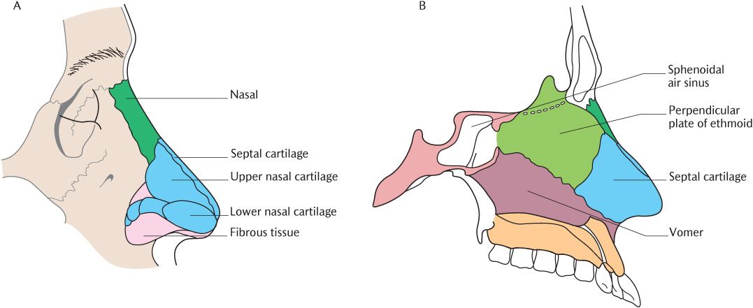
Fig. 27.2 The skeleton of the external nose. A) External view; B) View of the nasal septum.
The maxilla and the labyrinth of the ethmoid are the main contributors to the lateral wall which is irregular due to three medially projecting folds of bone, the nasal conchae. As Figure 27.1 illustrates, the ethmoid forms the superior portion down to, and including, the middle concha and the maxilla forms the lower part. The maxillary contribution to the nasal cavity is described below. The palatine bone forms the posterior part of the lateral wall. The palatine bone consists of horizontal and perpendicular plates arranged at right angles to each other like a letter L in frontal view. The horizontal plate forms the posterior part of the bony palate. As shown in Figure 27.3, the perpendicular plate articulates with the roughened posterior area of the medial surface of the maxilla to form the posterior part of the lateral nasal wall and covers over the posterior part of the maxillary hiatus, a large defect in the lateral nasal wall leading into the maxillary air sinus. In Figure 27.3, you can see that some of the inferior wall is overlain by the separate inferior nasal concha, a scroll-like plate of bone projecting into the nasal cavity. Figure 27.3 also shows the minor contributions of the nasal and lacrimal bones.
As shown in Figure 27.1, the conchae incompletely divide the nasal cavity into three horizontal passages—the superior meatus between the superior and middle conchae, the middle meatus between the middle and inferior conchae, and the inferior meatus between the inferior concha and the palate. The area of the lateral wall above the superior concha is termed the sphenoethmoidal recess because it forms a recess under those two bones.
The nasal surface of an isolated maxilla is shown in Figure 27.4; the shaded areas indicate where the maxilla articulates with other bones. Identify the prominent frontal process and follow it down to the smoothly curved medial surface. The upper posterior area is roughened where it articulates in a complex manner with the perpendicular plate of the palatine, ethmoid labyrinth, inferior concha, and lacrimal bones. The maxillary hiatus is the large opening into the maxillary sinus in this area, but is only visible on the maxilla in isolation. When the maxilla is articulated with the bones listed above, the maxillary hiatus is reduced to a single opening on the lateral wall between the superior and inferior conchae; sometimes there are a number of small openings.
The formation of the nasolacrimal groove in the lateral nasal wall is described in
Stay updated, free dental videos. Join our Telegram channel
VIDEdental - Online dental courses


