2: General Embryology

General Embryology

This chapter provides basic general embryology information needed to explain the development of the head, particularly the structures in and around the mouth. It supplies a background for understanding (1) the origins of the tissues associated with facial and dental development and (2) the cause of many congenital defects manifest in these tissues.

Germ Cell Formation and Fertilization

The human somatic (body) cell contains 46 chromosomes, 46 being the diploid number for the cell. Two of these are sex chromosomes; the remaining are autosomes. Each chromosome is paired so that every cell has 22 homologous sets of paired autosomes, with one sex chromosome derived from the mother and one from the father. The sex chromosomes, designated X and Y, are paired as XX in the female and XY in the male.

Fertilization is the fusion of male and female germ cells (the spermatozoa and ova, collectively called gametes) to form a zygote, which commences the formation of a new individual. Germ cells are required to have half as many chromosomes (the haploid number), so that on fertilization the original complement of 46 chromosomes will be reestablished in the new somatic cell. The process that produces germ cells with half the number of chromosomes of the somatic cell is called meiosis. Mitosis describes the division of somatic cells.

Before mitotic cell division begins, DNA is first replicated during the synthetic (S) phase of the cell cycle so that the amount of DNA is doubled to a value known as tetraploid (4 times the amount of DNA found in the germ cell). During mitosis the chromosomes containing this tetraploid amount of DNA are split and distributed equally between the two resulting cells; thus both daughter cells have a diploid DNA quantity and chromosome number, which duplicates the parent cell exactly.

Meiosis, by contrast, involves two sets of cell divisions occurring in quick succession. Before the first division, DNA is replicated to the tetraploid value (as in mitosis). In the first division the number of chromosomes is halved, and each daughter cell contains a diploid amount of DNA. The second division involves the splitting and separation of the chromosomes resulting in four cells; thus the final composition of each cell is haploid with respect to its DNA value and its chromosome number.

Meiosis is discussed in this textbook because the process occasionally malfunctions by producing zygotes with an abnormal number of chromosomes and individuals with congenital defects that sometimes affect the mouth and teeth. For example, an abnormal number of chromosomes can result from the failure to separate of a homologous chromosome pair during meiosis, so that the daughter cells contain 24 or 22 chromosomes. If, on fertilization, a gamete containing 24 chromosomes fuses with a normal gamete (containing 23), the resulting zygote will possess 47 chromosomes; one homologous pair has a third component. Thus the cells are trisomic for a given pair of chromosomes. If one member of the homologous chromosome pair is missing, a rare condition known as monosomy prevails. The best known example of trisomy is Down syndrome, or trisomy 21. Among features of Down syndrome are facial clefts, a shortened palate, a protruding and fissured tongue, and delayed eruption of teeth.

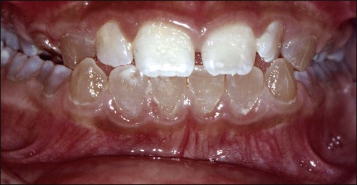
Approximately 10% of all human malformations are caused by an alteration in a single gene. Such alterations are transmitted in several ways, of which two are of special importance. First, if the malformation results from autosomal dominant inheritance, the affected gene generally is inherited from only one parent. The trait usually appears in every generation and can be transmitted by the affected parent to statistically half of the children. Examples of autosomal dominant conditions include achondroplasia, cleidocranial dysostosis, osteogenesis imperfecta, and dentinogenesis imperfecta; the latter two conditions result in abnormal formation of the dental hard tissues. Dentinogenesis imperfecta (Figure 2-1) arises from a mutation in the dentin sialophosphoprotein gene. Second, when the malformation is due to autosomal recessive inheritance, the abnormal gene can express itself only when it is received from both parents. Examples include chondroectodermal dysplasia, some cases of microcephaly, and cystic fibrosis.

All of these conditions are examples of abnormalities in the genetic makeup or genotype of the individual and are classified as genetic defects. The expression of the genotype is affected by the environment in which the embryo develops, and the final outcome of development is termed the phenotype. Adverse factors in the environment can result in excessive deviation from a functional and accepted norm; the outcome is described as a congenital defect. Teratology is the study of such developmental defects.

Prenatal Development

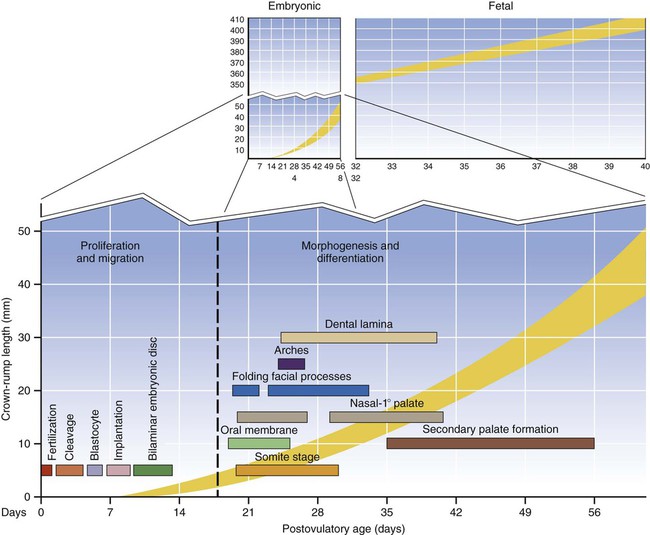
Prenatal development is divided into three successive phases. The first two, when combined, constitute the embryonic stage, and the third is the fetal stage. The forming individual is described as an embryo or fetus depending on its developmental stage.

The first phase begins at fertilization and spans the first 4 weeks or so of development. This phase involves largely cellular proliferation and migration, with some differentiation of cell populations. Few congenital defects result from this period of development because, if the perturbation is severe, the embryo is lost.

The second phase spans the next 4 weeks of development and is characterized largely by the differentiation of all major external and internal structures (morphogenesis). The second phase is a particularly vulnerable period for the embryo because it involves many intricate embryologic processes; during this period, many recognized congenital defects develop.

From the end of the second phase to term, further development is largely a matter of growth and maturation, and the embryo now is called a fetus (Figure 2-2).

Induction, Competence, and Differentiation

Patterning is key in development from the initial axial (head-to-tail) specification of the embryo through its segmentation and ultimately to the development of the dentition. Patterning is a spatial and temporal event as exemplified by regional development of incisors, canines, premolars, and molars, which occurs at different times and involves the classical processes of induction, competence, and differentiation.

All the cells of an individual stem from the zygote. Clearly, they have differentiated somehow into populations that have assumed particular functions, shapes, and rates of turnover. The process that initiates differentiation is induction; an inducer is the agent that provides cells with the signal to enter this process. Furthermore, each compartment of cells must be competent to respond to the induction process. Evidence suggests that over time, populations of embryonic cells vary their competence from no response to maximum response and then back to no response. In other words, windows of competence of varying duration exist for different populations of cells. The concepts of induction, competence, and differentiation apply in the development of the tooth and its supporting tissues.

Using probes composed of specific nucleic acid sequences, recombinant DNA technology can identify not only specific genes but also whether genes are transcriptionally active. By using antibodies for specific proteins, immunohistochemistry provides precise identification and localization of molecules within tissues and cells. These two technologies have led to the recognition of homeobox genes and growth factors, both of which play crucial roles in development.

All homeobox genes contain a similar region of 180 nucleotide base pairs (the homeobox) and function by producing proteins (transcription factors) that bind to the DNA of other downstream genes, thereby regulating their expression. By knocking out such genes or by switching them on, it has been shown that they play a fundamental role in patterning. Furthermore, combinations of differing homeobox genes provide codes or sets of assembly rules to regulate development; one such code is involved in dental development (see

Stay updated, free dental videos. Join our Telegram channel

Jan 5, 2015 | Posted by in General Dentistry | Comments Off on 2: General Embryology

VIDEdental - Online dental courses
Get VIDEdental app for watching clinical videos