10: Physiologic Tooth Movement: Eruption and Shedding

Physiologic Tooth Movement

Eruption and Shedding

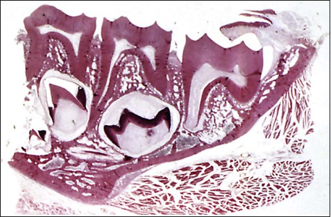
The jaws of an infant can accommodate only a few small teeth. Because teeth, when formed, cannot increase in size, the larger jaws of the adult require not only more but also bigger teeth. This accommodation is accomplished with two dentitions. The first is the deciduous or primary dentition, and the second is the permanent or secondary dentition (Figures 10-1 and 10-2).

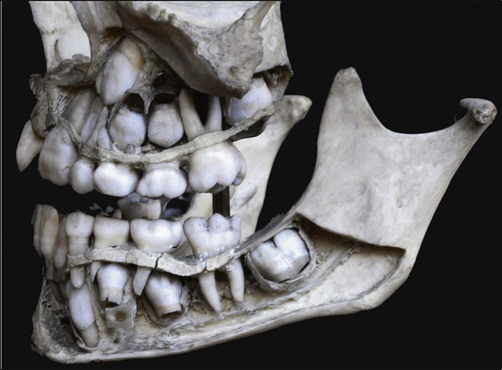
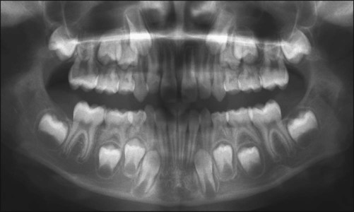
The early development of teeth has been described already, and the point has been made that the teeth develop within the tissues of the jaw (Figure 10-3). For teeth to become functional, considerable movement is required to bring them into the occlusal plane. The movements teeth make are complex and may be described in general terms as follows:

Superimposed on these movements is a progression from primary to permanent dentition, involving the shedding (or exfoliation) of the deciduous dentition. Although this categorization of tooth movement is convenient for descriptive purposes, what is being described is a complex series of events occurring in a continuous process to move the tooth in three-dimensional space.

Preeruptive Tooth Movement

When the deciduous tooth germs first differentiate, they are extremely small, and a good deal of space is available for them in the developing jaw. Because they grow rapidly, however, they become crowded. A lengthening of the jaws, which permits the deciduous second molar tooth germs to move backward and the anterior germs to move forward gradually, alleviates this crowding. At the same time the tooth germs are moving bodily outward and upward (or downward, as the case may be) with the increasing length and width and height of the jaws.

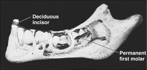
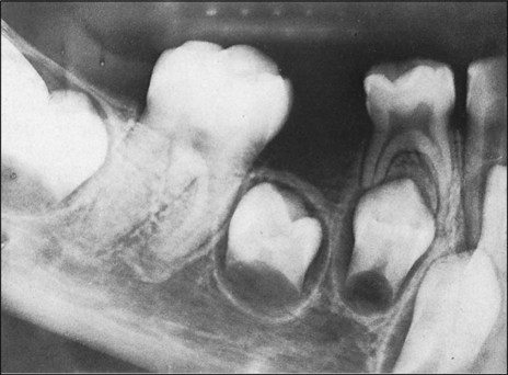
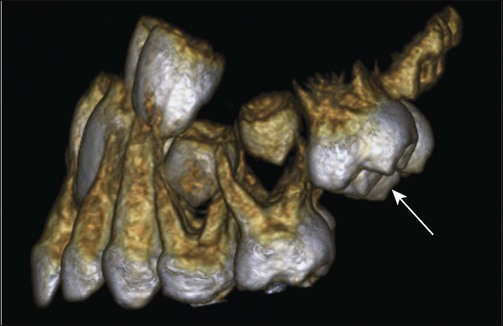
The origin of the successional permanent teeth was described in Chapter 5. Those tooth germs develop on the lingual aspect of their deciduous predecessors, in the same bony crypt. From this position they shift considerably as the jaws develop. For example, the incisors and canines eventually occupy a position, in their own bony crypts, on the lingual side of the roots of their deciduous predecessors, and the premolar tooth germs, also in their own crypts, finally are positioned between the divergent roots of the deciduous molars (Figures 10-4 and 10-5).

The permanent molar tooth germs, which have no predecessors, develop from the backward extension of the dental lamina. At first, little room is available in the jaws to accommodate these tooth germs. In the upper jaw the molar tooth germs develop first, with their occlusal surfaces facing distally, and then swing into position only when the maxilla has grown sufficiently to provide room for such movement (Figure 10-6). In the mandible the permanent molars develop with their axes showing a mesial inclination, which becomes vertical only when sufficient jaw growth has occurred.

These preeruptive movements of deciduous and permanent tooth germs place the teeth in a position within the jaw for eruptive movement. These preeruptive movements of teeth are a combination of two factors: (1) total bodily movement of the tooth germ and (2) growth in which one part of the tooth germ remains fixed while the rest continues to grow, leading to a change in the center of the tooth germ. This growth explains, for example, how the deciduous incisors maintain their position relative to the oral mucosa as the jaws increase in height.

Preeruptive movements occur in an intraosseous location and are reflected in the patterns of bony remodeling within the crypt wall. For example, during bodily movement in a mesial direction, bone resorption occurs on the mesial surface of the crypt wall, and bone deposition occurs on the distal wall as a filling-in process. During eccentric growth, only bony resorption occurs, thus altering the shape of the crypt to accommodate the altering shape of the tooth germ. Little is known about the mechanisms that determine preeruptive tooth movements, including whether remodeling of bone to position the bony crypt is important as a mechanism or merely represents an adaptive response.

Eruptive Tooth Movement

The mechanisms of eruption for deciduous and permanent teeth are similar, resulting in the axial or occlusal movement of the tooth from its developmental position within the jaw to its final functional position in the occlusal plane. The actual eruption of the tooth, when it breaks through the gum, is only one phase of eruption.

Histologic Features

Histologically, many changes occur in association with and for the accommodation of tooth eruption. The periodontal ligament (PDL) develops only after root formation has been initiated; when established, the PDL must be remodeled to accommodate continued eruptive tooth movement. The remodeling of PDL fiber bundles is achieved by the fibroblasts, which simultaneously synthesize and degrade the collagen fibrils as required across the entire extent of the ligament. Recall also that the fibroblast has a cytoskeleton, which enables it to contract. This contractility is a property of all fibroblasts but is especially well developed in PDL fibroblasts, which have been demonstrated to exert stronger contractile forces than, for example, gingival or skin fibroblasts. Ligament fibroblasts exhibit numerous contacts with one another of the adherens type and exhibit a close relationship to PDL collagen fiber bundles.

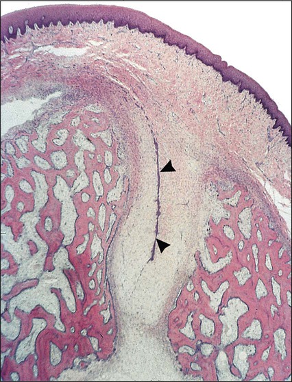
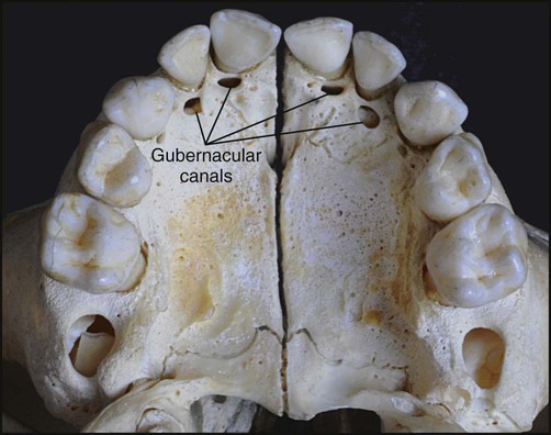
The architecture of the tissues in advance of erupting successional teeth differs from that found in advance of deciduous teeth. The fibrocellular follicle surrounding a successional tooth retains its connection with the lamina propria of the oral mucous membrane by means of a strand of fibrous tissue containing remnants of the dental lamina, known as the gubernacular cord. In a dried skull, holes can be identified in the jaws on the lingual aspects of the deciduous teeth. These holes, which once contained the gubernacular cords, are termed gubernacular canals (Figures 10-7 and 10-8). As the successional tooth erupts, its gubernacular canal is widened rapidly by local osteoclastic activity, delineating the eruptive pathway for the tooth. The rate of eruption depends on the phase of movement. During the intraosseous phase, the rate can attain 10 mm per day; it increases to about 75 mm per day once the tooth escapes from its bony crypt. This rate persists until the tooth reaches the occlusal plane, indicating that soft connective tissue provides little resistance to tooth movement.

When the erupting tooth appears in the oral cavity, it is subjected to environmental factors that help determine its final position in the dental arch. Muscle forces from the tongue, cheeks, and lips play on the tooth, as do the forces of contact of the erupting tooth with other erupted teeth. The childhood habit of thumb-sucking is an obvious example of environmental influence of tooth position.

Mechanisms of Eruptive Tooth Movement

Eruptive mechanisms are not understood fully yet, but it is generally believed that eruption is a multifactorial process in which cause and effect are difficult to separate. Numerous theories for tooth eruption have been proposed; among these, root elongation, alveolar bone remodeling, and to some extent, formation of the PDL provide the most plausible explanation for tooth eruption in human beings. An excellent critical review on the factors involved in tooth eruption has been written by Marks and Schroeder (see Recommended Reading).

Root Formation

At first glance, root formation appears to be an obvious cause of tooth eruption because it undoubtedly causes an overall increase in the length of the tooth that must be accommodated by the growth of the root into the bone of the jaw, by an increase in jaw height, or by the occlusal movement of the crown. Although the last movement is what occurs, it does not follow that root growth is responsible. Indeed, clinical observation, experimental studies, and histologic analysis argue strongly against such a conclusion. For example, if a continuously erupting tooth (e.g., the guinea pig molar) is prevented from erupting by being pinned to bone, root growth continues and is accommodated by resorption of some bone at the base of the socket and by a buckling of the newly formed root. This experiment yields two conclusions: that root growth produces a force and that this force is sufficient to produce bone resorption. Thus although root growth can produce a force, it cannot be translated into eruptive tooth movement unless some structure exists at the base of the tooth capable of withstanding this force; because no such structure exists, some other mechanism must move the tooth to accommodate root growth. The situation is substantiated further by the facts that rootless teeth erupt, that some teeth erupt a greater distance than the total length of their roots, and that teeth still will erupt after the completion of root formation.

In conclusion, root formation per se is not required for tooth eruption, although root formation, under certain circumstances, may accelerate tooth eruption. Depending on the rate at which the root elongates, the basal bone will resorb or form to maintain a proper relationship between the root and bone.

Bone Remodeling

Bone remodeling of the jaws has been linked to tooth eruption in that, as in the preeruptive phase, the inherent growth pattern of the mandible or maxilla supposedly moves teeth by the selective deposition and resorption of bone in the immediate neighborhood of the tooth. The strongest evidence in support of bone remodeling as a cause of tooth movement comes from a series of experiments in dogs. When the developing premolar is removed without disturbing the dental follicle, or if eruption is prevented by wiring the tooth germ down to the lower border of the mandible, an eruptive pathway still forms within the bone overlying the enucleated tooth as osteoclasts widen the gubernacular canal. If the dental follicle is removed, however, no eruptive pathway forms. Furthermore, if a metal or silicone replica replaces the tooth germ, and so long as the dental follicle is retained, the replica will erupt, with the formation of an eruptive pathway. These observations should be analyzed carefully. First, they clearly demonstrate that an eruptive pathway can form in bone without a developing and growing tooth). Second, they show that the dental follicle is involved. The conclusion cannot be drawn that the demonstration of an eruptive pathway forming within bone means that bony remodeling is responsible for tooth movement unless coincident bone deposition also can be demonstrated at the base of the crypt and prevention of such bone deposition can be shown to interfere with tooth eruption. Careful studies using tetracyclines as markers of bone deposition have shown that the predominant activity in the fundus of an alveolus in a number of species (including human beings) is bone resorption. In humans, for instance, the base of the crypt of the permanent first and third molars continually resorbs as these teeth erupt, although in the second premolar and molar, some bone deposition on the crypt floor occurs. In the case of the demonstrated eruption of an inert replica, one might think that only bony remodeling could bring this about, but as discussed next, evidence indicates that follicular tissue is responsible for this movement. In addition, some recent studies are showing that alveolar bone growth at the base of the crypt is required for molar tooth eruption in rat. Clearly, the intraosseous tooth eruption needs further attention. Irrespective of whether bone growth is a primary moving force, it is generally agreed that the dental follicle is needed for eruption to occur and that, as discussed next, it modulates bone remodeling.

Dental Follicle

Investigations indicate a pattern of cellular activity involving the reduced dental epithelium and the follicle associated with tooth eruption, which facilitates connective tissue degradation and bone resorption as the tooth erupts. In osteopetrotic animals, which lack colony-stimulating factor 1, a factor that stimulates differentiation of osteoclasts, eruption is prevented because no mechanism for bone removal exists. Local administration of this factor permits the differentiation of osteoclasts, and eruption occurs. The reduced enamel epithelium also secretes proteases, which assist in the breakdown of connective tissue to produce a path of least resistance. Expression of bone morphogenetic protein-6 in the dental follicle may also be essential for promoting alveolar bone growth at the base of the crypt.

It is believed that there is signaling between the reduced enamel epithelium and dental follicle. This signaling could explain the remarkable consistency of eruption times, for the enamel epithelium likely is programmed as part of its functional life cycle. Signaling also helps to explain why radicular follicle, which is not associated with reduced enamel epithelium, does not undergo degeneration but instead participates in the formation of the PDL.

Stay updated, free dental videos. Join our Telegram channel

Jan 5, 2015 | Posted by in General Dentistry | Comments Off on 10: Physiologic Tooth Movement: Eruption and Shedding

VIDEdental - Online dental courses

Get VIDEdental app for watching clinical videos