Differential Diagnosis of Oral Lesions and Developmental Anomalies
Developmental anomalies (Table 2-1, Figure 2-1)
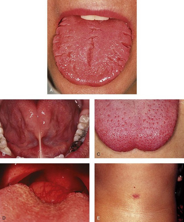
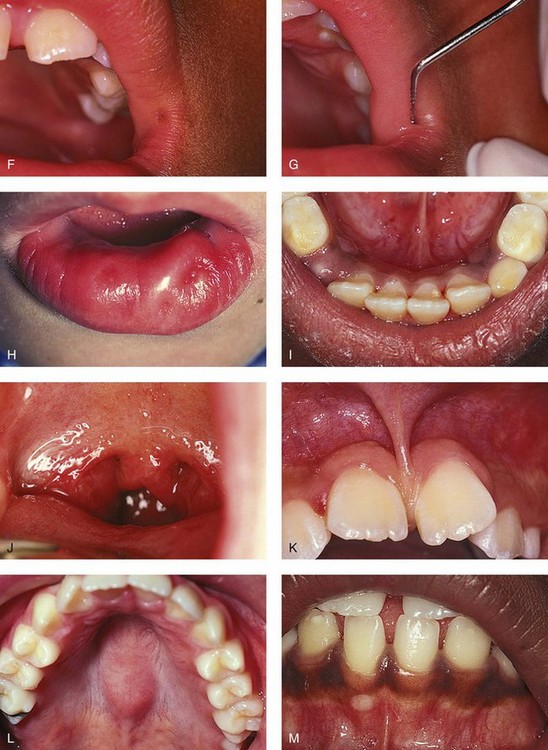
 FIGURE 2-1 Developmental anomalies.
A, Fissured tongue. B and C, Partial ankyloglossia with lingual frenum attachment at the tip of the tongue (B). Note the restricted mobility of the tongue with extension (C). D, Lingual thyroid of the midline base of the tongue. E, Thyroglossal duct cyst with sinus tract, midline neck. F and G, Commissural lip pit (F) with depth illustrated by periodontal probe (G). H, Paramedian lip pits. I, Retrocuspid papilla of the lingual mandibular gingiva. J, Bifid uvula. K, Hyperplastic maxillary labial frenum. L, Torus palatinus of the midline hard palate. M, Small exostosis of the anterior mandibular alveolus, facial aspect. (D courtesy Dr. G. E. Lilly, University of Iowa College of Dentistry.)
White soft tissue lesions (Table 2-2, Figure 2-2)
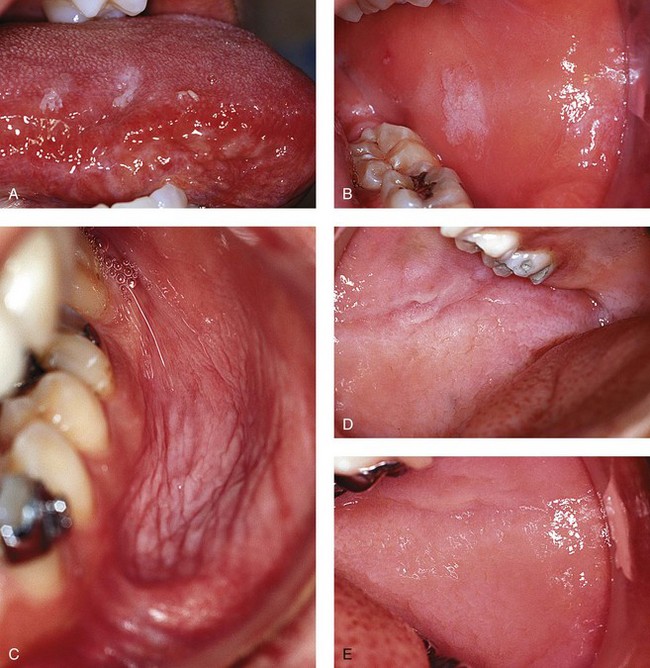
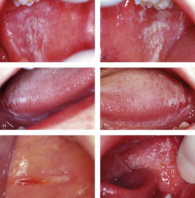
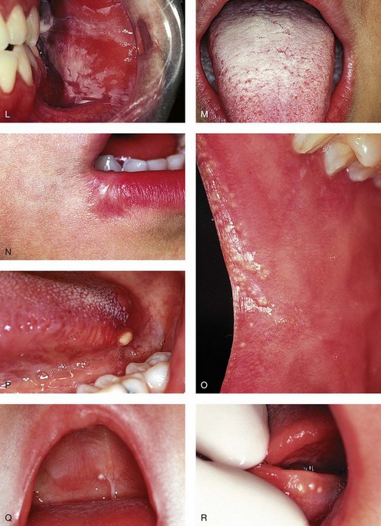
 FIGURE 2-2 White soft tissue lesions.
A and B, Frictional keratosis of the lateral tongue (A) and buccal mucosa (B) from chronic biting of the tissues. C, Smokeless tobacco keratosis of the posterior mandibular vestibule. D and E, Leukoedema of the buccal mucosa, bilaterally. F to I, White sponge nevus of the buccal mucosa (F and G) and lateral tongue (H and I). J, Ulcerated linea alba from aggressive sucking habit. K, Pseudomembranous candidiasis of the buccal mucosa. L, Chemical burn from overuse of a topical anesthetic. M, Coated tongue in a child who is mouth breathing. N, Fan-shaped scar at the corners of the mouth due to an electrical burn. O, Cluster of Fordyce granules of the anterior buccal mucosa. P, Oral lymphoepithelial cyst of the posterior lateral tongue. Q, Single palatal cyst of the newborn on the midline hard palate. R, Cluster of gingival cysts of the newborn on the mandibular alveolar mucosa.
Stay updated, free dental videos. Join our Telegram channel

VIDEdental - Online dental courses


 TABLE 2-1
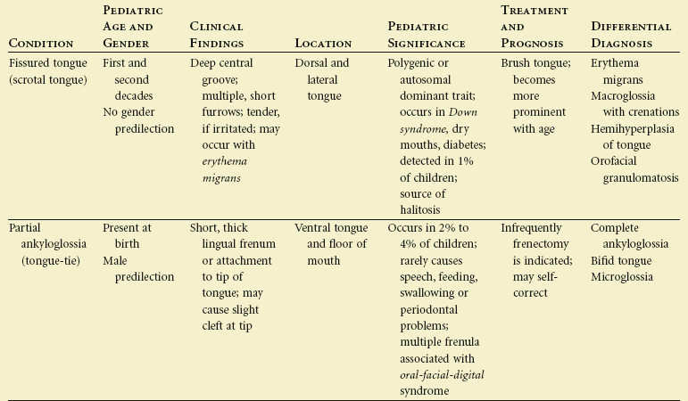
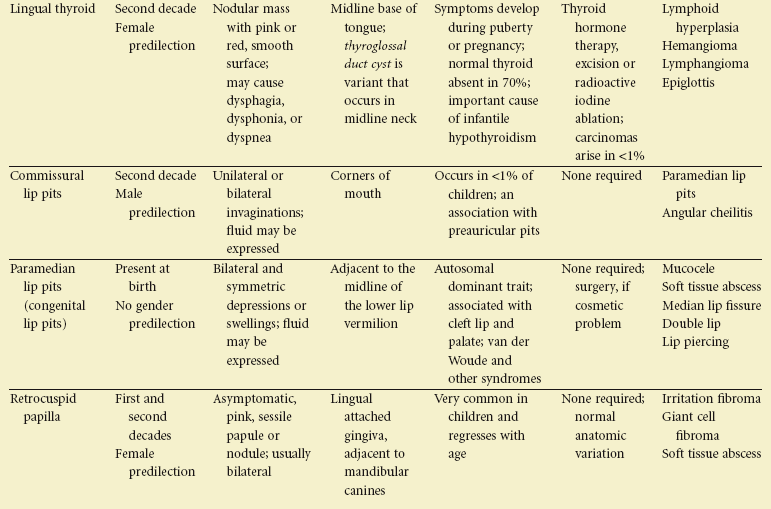
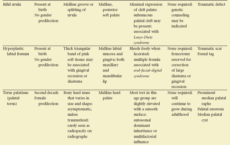
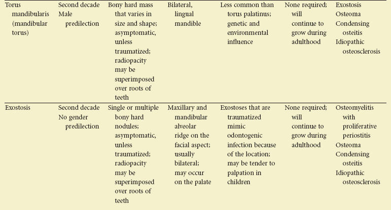
 TABLE 2-2
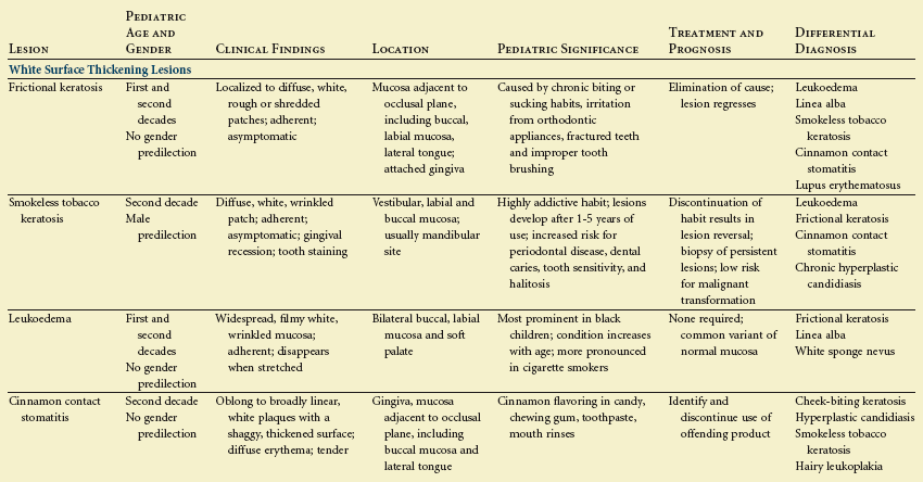
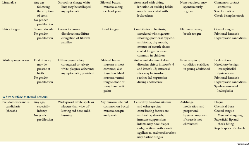
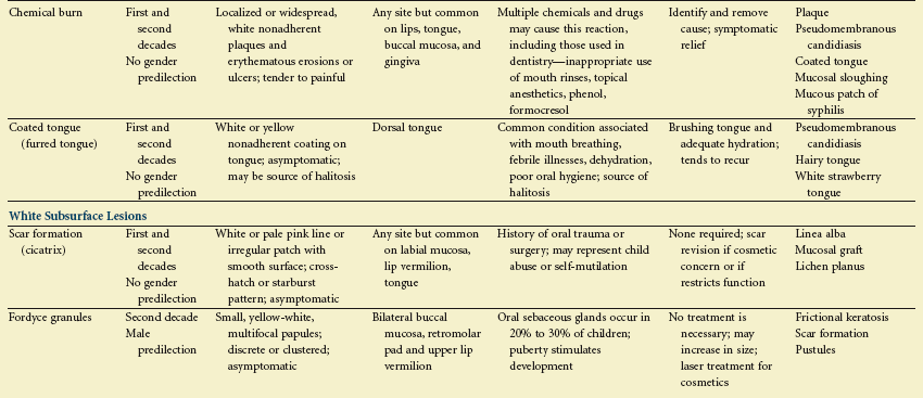
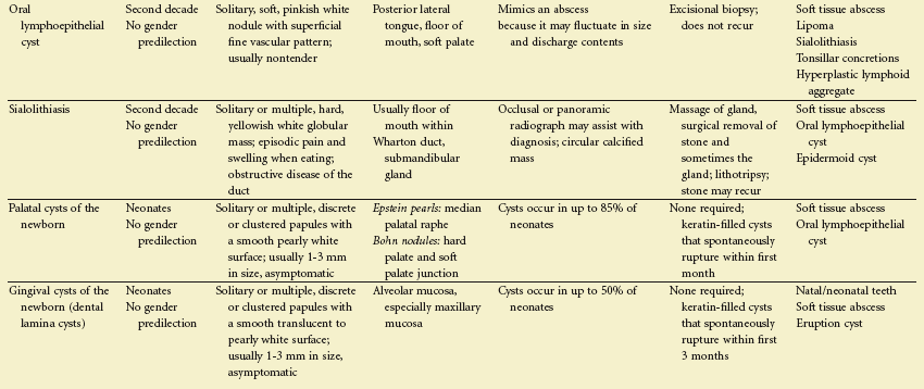
 TABLE 2-3
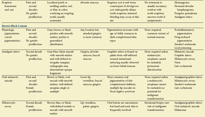
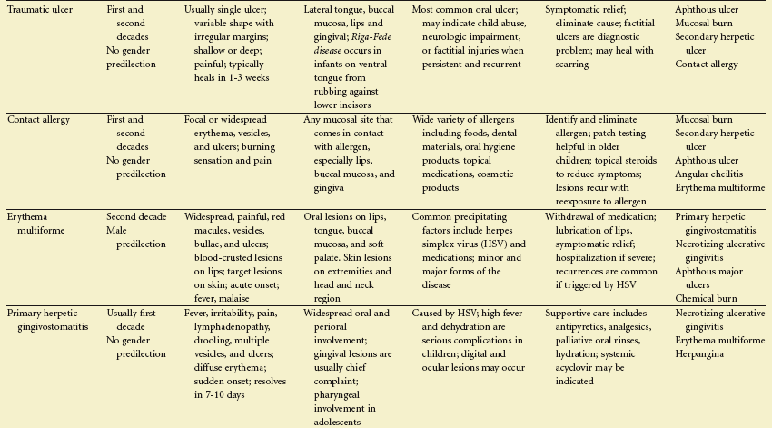
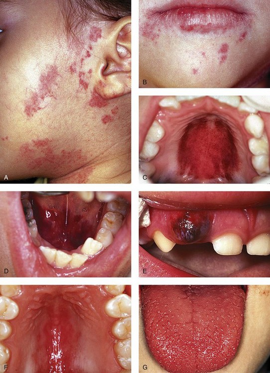
 FIGURE 2-3