12 – Smile Osteotomy

e9780867155457_i0584.jpg

CHAPTER 12

Smile Osteotomy

Ole T. Jensen, DDS, MS

After scientific discovery, nature’s enigmatic smile.

—O. Brekne

The atrophic, partially edentulous posterior segment of the mandible with a relatively prominent endosseous nerve location and uncertain prognosis for vertical onlay bone grafting can be treated with an osteoperiosteal flap that is designated the smile osteotomy. The smile osteotomy is a sandwich graft with a basal cut curved like a smile; the lowest part of the osteotomy is located 2 mm above the nerve, at the midpoint of the osteotomy, before the cut is tapered crestally both anteriorly and posteriorly.1

e9780867155457_i0585.jpg Rationale

The smile osteotomy is indicated when there is less than 8 mm of bone above the inferior alveolar nerve, a relatively deep alveolar plane, and the prospect of a poor crown-implant ratio.2 Nerve transposition, block or particulate onlay grafting, tent pole–guided bone regeneration (GBR), or even high-profile implants used to obtain tent pole–GBR are the alternatives.3–6 A failure of any of these various surgical approaches can leave the area in a worse condition than was found initially. The reported complication rate of nerve transposition surgery is about 5%.7 The complication rate is from 5% to 40% for block grafting and from 5% to 20% for GBR.8,9 The surgeon must decide what he or she considers an acceptable complication rate. When morbidity approaches 1 in 10 patients, this may give the surgeon reason enough to consider an alternative option.

The technique of nerve transposition, although often completed with minimal complications, still results in a significant incidence of paresthesia, and permanent painful dysesthesia is not infrequent.3,10 This procedure probably should seldom be performed electively because dysesthesia can result even in the absence of any recognizable trauma to the neural bundle. Also, individuals in older age groups may have less tolerance for paresthesia.11 It is probably best to avoid this highly technical procedure, even if done with piezoelectric surgery for relatively short manipulations of less than 10 mm in length, unless there is no alternative, such as in tumor ablations.12

Onlay block grafting has been tried with allograft blocks,3 mandibular monocortical blocks, cranial blocks, and iliac crest corticocancellous blocks.13–16 All of these procedures have been known to work and, at times, not to work. The monocortical block graft harvested from the ramus or chin has been shown to have about a 5% rate of complete graft loss early and up to a 40% rate of partial loss later, usually due to resorption and/or poor consolidation.8 The use of corticocancellous block from the ilium is much more conducive to consolidation, but vertical resorption can be 50% of volume or greater by 3 years.16

The use of allograft blocks is not well reported, but the material has the disadvantage of not being autogenous bone; therefore, allografts would be expected to exhibit less consolidation and greater dehiscence than found in autografts, although the resorption rate may be less pronounced. 17

The use of GBR or particulate grafting is problematic for vertical augmentation of greater than 5 mm, especially at interdental sites, because membrane exposure or graft infection may result. When this occurs, the site is at risk for complete graft loss.9

When high-profile implants are placed as tent poles in a membrane technique, there is a risk that osseointegration will occur despite complete graft loss, leaving exposed screw threads.6

Thus, all the aforementioned procedures in the posterior mandible have the potential for significant complications. The sandwich bone graft, on the other hand, has been demonstrated to have almost none of these problems; that is, it has a very low rate of infection or graft loss, exhibits excellent consolidation, and results in a stable long-term alveolar morphology.1

Perhaps the only reason to avoid this procedure is a technical one. However, most oral and maxillofacial surgeons familiar with orthognathic surgery can easily master the procedure.1

The problem with performing a sandwich osteotomy in the posterior mandible is one of access. However, using the principles learned with anterior segmental surgery, the surgeon can treat this area of the mouth just as well as the anterior segment. The surgeon must be familiar with both piezoelectric surgery and oscillating saw techniques. Once the surgeon has mastered the technical aspects of the procedure, he or she will develop the surgical judgment to determine when and when not to perform the procedure.

e9780867155457_i0587.jpg Surgical Technique

Following administration of local and intravenous anesthesia, a mouth prop is used to open the mouth on the opposite side to establish surgical access. A buccal vestibular incision is carried out about 5 mm below the attached gingiva along the length of the edentulous space to the retromolar pad; the mental foramen and associated nerve bundle must be carefully avoided (Fig 12-1a). The intraosseous nerve location is identified by using a computed tomography (CT) scan preoperatively to help design the osteotomy so that it can be performed without reflecting the flap too far crestally. (The osteotomy can be performed without any reflection of mucoperiosteum toward the crest.) The closest to the nerve that the osteotomy gets inferiorly should be in the center of the segment—about 2 mm above the nerve—before the cut tapers to the crest posteriorly and anteriorly. The anterior cut should stay about 2 mm from the most distal tooth root.

e9780867155457_i0588.jpg

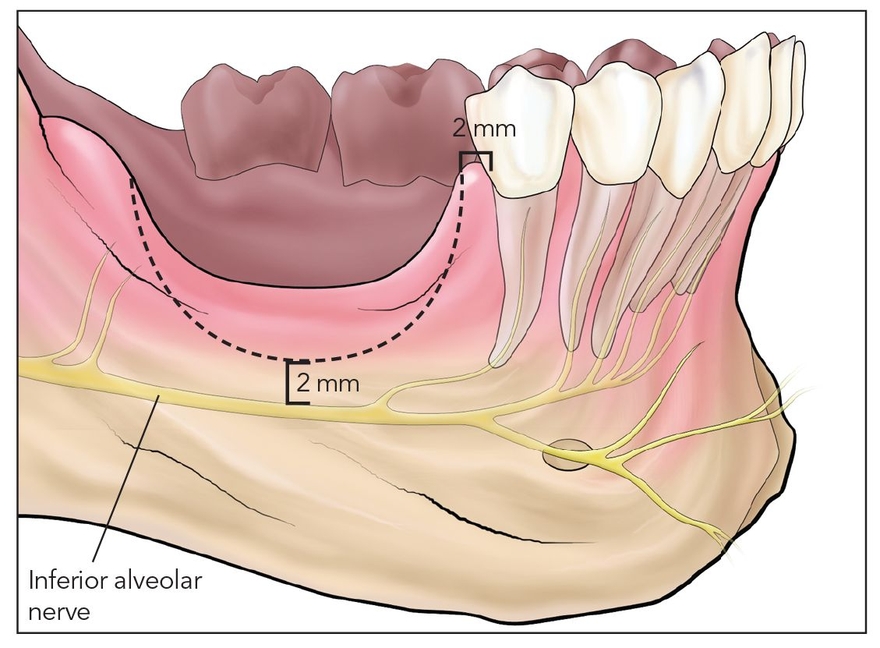
Fig 12-1a The posterior atrophic mandible often dips down vertically near the inferior alveolar nerve. A segmental osteotomy (dotted line) is designed in a curved shape, termed the smile osteotomy, that minimizes the proximity of the bony cut to the nerve.

e9780867155457_i0589.jpg

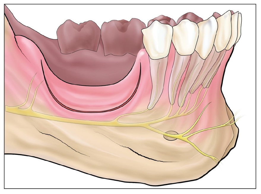
Fig 12-1b The segment is cut at least 2 mm above the nerve using piezoelectric surgery (shallow) and an oscillating saw (deep).

e9780867155457_i0590.jpg

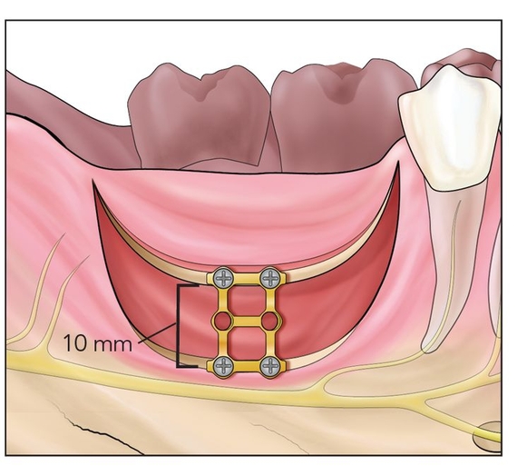
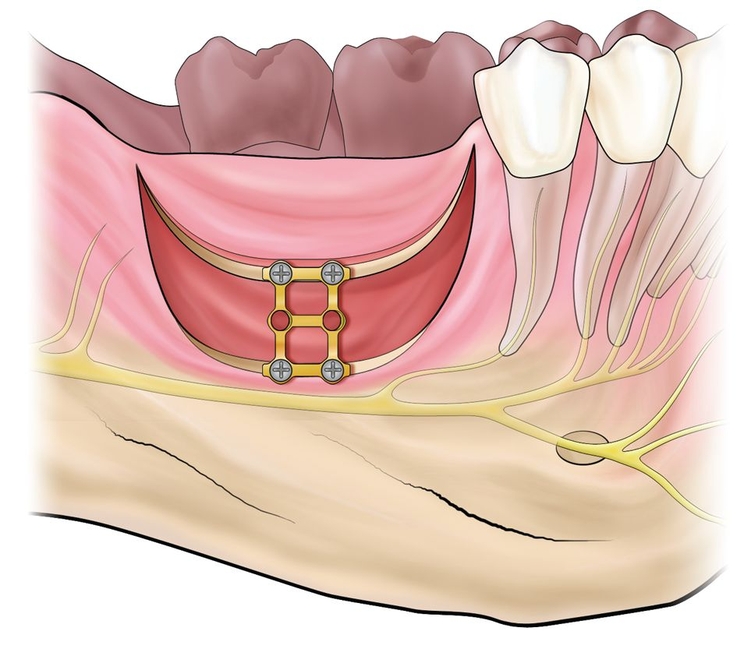
Fig 12-1c The segment is freed with an osteotome and elevated 5 to 10 mm vertically. A bone plate is placed to establish vertical height at the alveolar plane.

e9780867155457_i0591.jpg

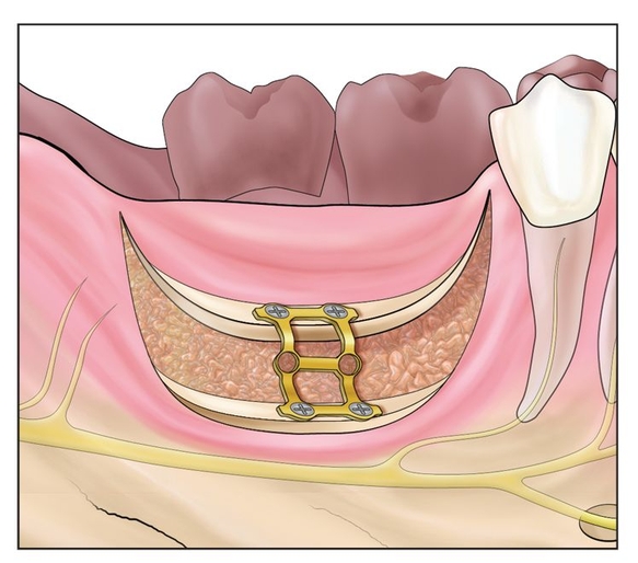
Fig 12-1d The bone plate is torqued to bring the alveolus into axial alignment and then interpositionally bone grafted with autograft or bone morphogenetic protein 2.

e9780867155457_i0592.jpg

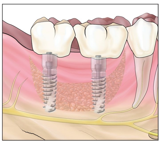
Fig 12-1e After 4 months of healing, implants are placed and later restored.

The initial smile-shaped cut can best be made with a piezoelectric knife to a depth of about 5 mm. The use of piezoelectric surgery is a good idea to delineate the osteotomy cut and to help avoid nerve injury. Once the smile-shaped lateral osteotomy cut is delineated, a sagittal saw is used to cut deeper to free the lingual plate (Fig 12-1b). The piezoelectric knife may not be well irrigated and cooled sufficiently to allow osseous cuts at />

Only gold members can continue reading. Log In or Register to continue

Stay updated, free dental videos. Join our Telegram channel

Jan 8, 2015 | Posted by in Implantology | Comments Off on 12 – Smile Osteotomy

VIDEdental - Online dental courses

Get VIDEdental app for watching clinical videos