10 The Permanent Mandibular Premolars
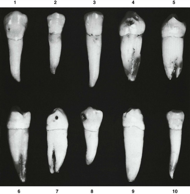
The first premolar has a large buccal cusp, which is long and well formed, with a small, nonfunctioning lingual cusp that in some specimens is no longer than the cingulum found on some maxillary canines (see Figure 10-10, 3 and 8; and Figure 10-12, 4 and 7). The second premolar has three well-formed cusps in most cases, one large buccal cusp and two smaller lingual cusps. The form of both mandibular premolars fails to conform to the implications of the term bicuspid, a term that implies two functioning cusps.
Mandibular First Premolar
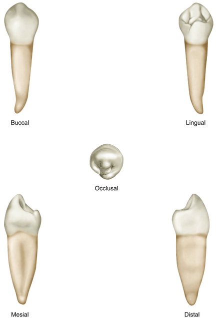
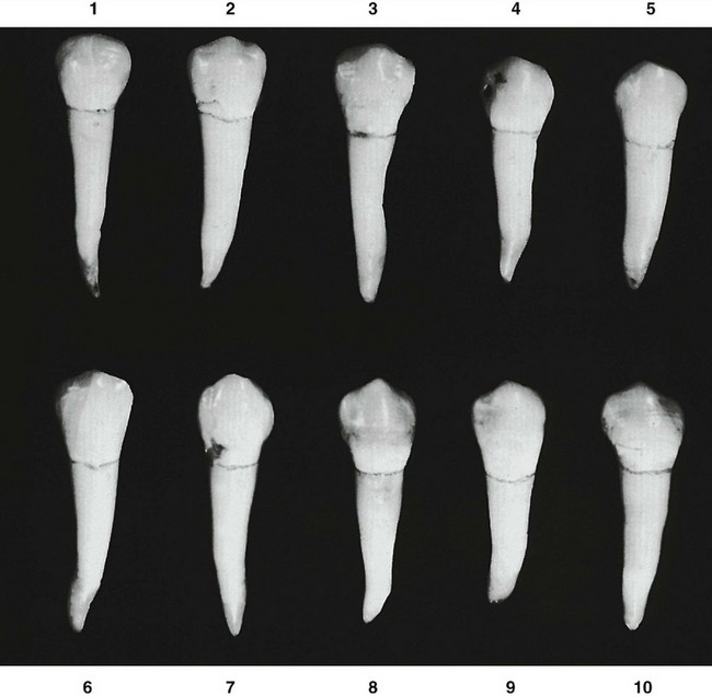
Figures 10-1 through 10-12 illustrate the mandibular first premolar from all aspects. The mandibular first premolar is the fourth tooth from the median line and the first posterior tooth in the mandible. This tooth is situated between the canine and second premolar and has some characteristics common to each of them.
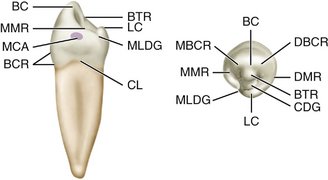
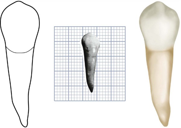
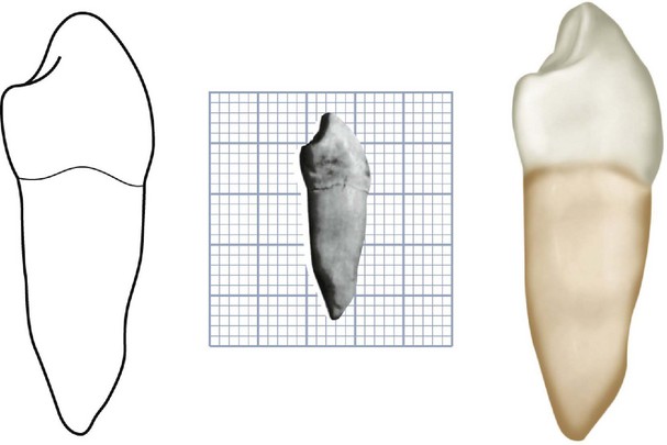
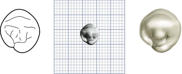
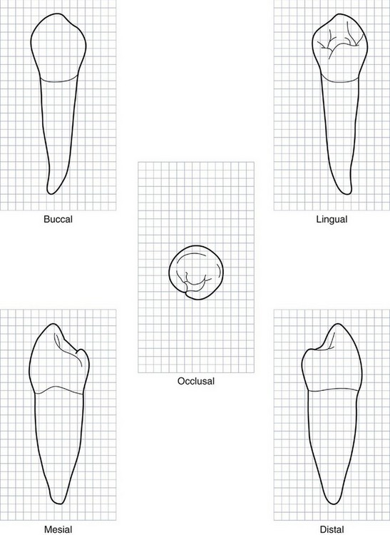
Figure 10-7 Mandibular right first premolar. Graph outlines of five aspects are shown. (Grid = 1 sq mm.)
The characteristics that resemble those of the mandibular canine are as follows:
The characteristics that resemble those of the second mandibular premolar are as follows:
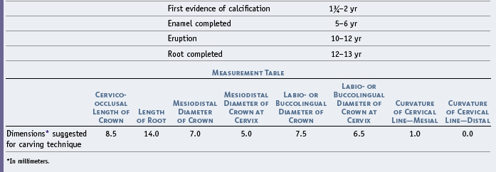
Although the root of the mandibular first premolar is shorter generally than that of the mandibular second premolar, it is closer to the length of the second premolar root than it is to that of the mandibular canine (Table 10-1).
DETAILED DESCRIPTION OF THE MANDIBULAR FIRST PREMOLAR FROM ALL ASPECTS
Buccal Aspect
From the buccal aspect, the form of the mandibular first premolar crown is nearly symmetrical bilaterally (see Figures 10-2, 10-7, 10-8, and 10-9). The middle buccal lobe is well developed, which results in a large, pointed buccal cusp. The mesial cusp ridge is shorter than the distal cusp ridge.
From the buccal aspect, the crown is roughly trapezoidal (see Figure 4-16, C). The cervical margin is represented by the shortest of the uneven sides.
The tip of the buccal cusp is pointed and, in most cases, is located a little mesial to the center of the crown buccally (see Figure 10-9, 3, 7, 8, and 9). The mandibular canine has the same characteristic to a greater degree.
The development of the middle buccal lobe is outstanding, ending in a pointed buccal cusp. Developmental depressions are often seen between the three lobes (see Figure 10-9, 2, 3, 8, and 10).
The continuous ridge from the cervical margin to the cusp tip is called the buccal ridge.
Lingual Aspect
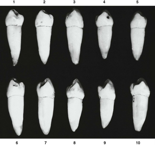
The crown of the mandibular first premolar tapers toward the lingual, since the lingual measurement mesiodistally is less than that buccally. The lingual cusp is always small (see Figures 10-3, 10-7, and 10-8). The major portion of the crown is made up of the middle buccal lobe (see Figure 10-11). This makes it resemble the canine.
Stay updated, free dental videos. Join our Telegram channel
VIDEdental - Online dental courses


