Panoramic Imaging
Sanjay M. Mallya and Alan G. Lurie
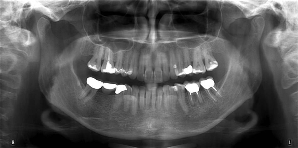
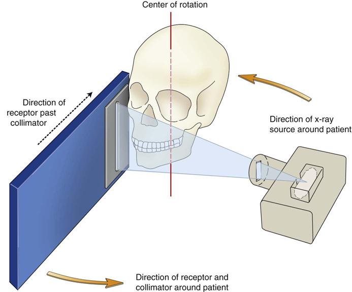
Panoramic imaging (also called pantomography) is a technique for producing a single image of the facial structures that includes both the maxillary and the mandibular dental arches and their supporting structures (Fig. 10-1). This technique produces a tomographic image in that it selectively images a specific body layer. In panoramic radiography, an x-ray source and an image receptor rotate around the patient’s head (Fig. 10-2) and create a curved focal trough, a zone in which the included objects are displayed clearly. Objects in front of or behind this focal trough are blurred and largely not seen. The panoramic machine thus creates a focal trough through the dentition and adjacent structures.
Panoramic images are most useful clinically for diagnostic problems requiring broad coverage of the jaws (Box 10-1). Common examples include evaluation of trauma including jaw fractures, location of third molars, extensive dental or osseous disease, known or suspected large lesions, tooth development and eruption (especially in the mixed dentition), retained teeth or root tips (in edentulous patients), temporomandibular joint (TMJ) pain, and developmental anomalies. Panoramic imaging is often used as the initial evaluation image that can provide the required insight or assist in determining the need for other projections. Panoramic images are also useful for patients who do not tolerate intraoral procedures well.
The main disadvantage of panoramic radiology is that the image does not display the fine anatomic detail available on intraoral periapical radiographs. Thus, it is not as useful as periapical radiography for detecting small carious lesions, fine structure of the marginal periodontium, or periapical disease. The proximal surfaces of premolars also typically overlap. The availability of a panoramic radiograph for an adult patient often does not preclude the need for intraoral films for the diagnoses of most commonly encountered dental diseases. When a full-mouth series of radiographs is available for a patient requiring only general dental care, typically little or no additional useful information is gained from a simultaneous panoramic examination. Other problems associated with panoramic radiography include unequal magnification and geometric distortion across the image. Occasionally, the presence of overlapping structures, such as the cervical spine, can hide odontogenic lesions, particularly in the incisor regions. Clinically important objects may be situated outside the focal trough and may appear distorted or not be seen at all.
Principles of Panoramic Image Formation
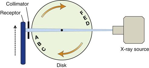
Paatero and, independently, Numata were the first to describe the principles of panoramic radiography. Figure 10-2 shows a schematic view of the relationships between the x-ray source, the patient, the secondary collimator, and the image receptor during panoramic image formation. The following illustrations explain the formation of the focal trough in a panoramic machine. Imagine an assembly containing a disk with upright physical objects (represented by letters) and an image receptor (Fig. 10-3). The receptor travels upward through the beam at the same speed as objects A through C rotate through the beam. A lead collimator in the shape of a slit located at the x-ray source limits the x-rays to a narrow vertical beam. Another collimator between the objects and the image receptor reduces scattered radiation from the objects to the image receptor. Consider first radiopaque objects A through C. As the disk rotates, their radiographic images are recorded sharply on the receptor that also moves through the beam at the same direction and speed. The spatial relationship of the shadows of these objects correctly represents the relationship of the actual objects. Because the source-receptor distance is constant and the object-receptor distance is the same for each object, all objects are magnified equally. Now consider objects D through F. They are located on the opposite side of the disk, between the x-ray source and the center of rotation of the disk. These objects move in the opposite direction of the receptor, so their shadows are reversed on the receptor. Because these objects are much closer to the x-ray source, their images are greatly magnified.
Figure 10-4 shows that the same relationship between the rotating receptor and objects can be achieved if the disk is held stationary but the x-ray source and the receptor are rotated around the center of rotation in the disk. The x-ray beam still passes through the center of the disk and sequentially through objects A through C. Similarly, the receptor is still moved through the beam and at the same rate as the beam passes through A through C. In this situation, as before, objects A through C move through the x-ray beam in the same direction and at the same rate as the receptor. Objects D through F continue to be blurred, just as before.
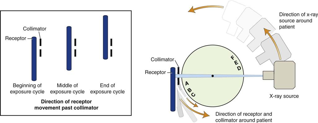
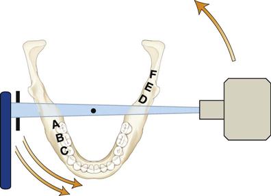
Figure 10-5 shows that a patient may replace the disk and objects A through F represent teeth and surrounding bone. The illustration demonstrates the positions of the x-ray source and the receptor early in an exposure cycle. The center of rotation is located off to the side of the arch, away from the objects being imaged. The rate of movement of the receptor is regulated to be the same as that of the x-ray beam sweeping through the dentoalveolar structures on the side of the patient nearest the receptor. Structures on the opposite side of the patient (near the x-ray tube) are distorted and appear out of focus because the x-ray beam sweeps through them in the direction opposite that in which the image receptor is moving. In addition, structures near the x-ray source are so magnified (and their borders so blurred) that they are not seen as discrete images on the resultant image. These structures appear only as diffuse phantom or ghost images. Because of both of these circumstances, only structures near the receptor are usefully captured on the resultant image.
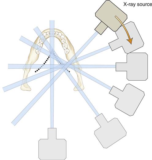
Contemporary panoramic machines use a continuously moving center of rotation rather than multiple fixed locations (Fig. 10-6). This feature optimizes the shape of the focal trough to reveal best the teeth and supporting bone. This center of rotation is initially near the lingual surface of the right body of the mandible when the left TMJ region is being imaged. The rotation center moves anteriorly along an arc that ends just lingual to the symphysis of the mandible when the midline is imaged. The arc is reversed as the opposite side of the jaws is imaged.
This basic principle of image formation remains the same, regardless of the type of detector used to record the image. In the case where the receptor is a charge-coupled device (CCD) array, the film is replaced by a two-dimensional CCD array. Each column of the array is read out to construct the image. The key is to read out the columns at the same rate as an imaginary moving film would move past the array. The CCD array is read out continuously as the x-ray source and receptor travel around the patient. The resulting geometric projection characteristics are the same as if film or a photostimulable phosphor plate (PSP) had been used; this holds true for geometric distortions such as magnification and elongation, the presence of ghost images, superimposition of the cervical spine over midline structures, overlap of teeth, and left-right size variations from lack of proper positioning of the patient’s sagittal plane in the instrument.
Focal Trough
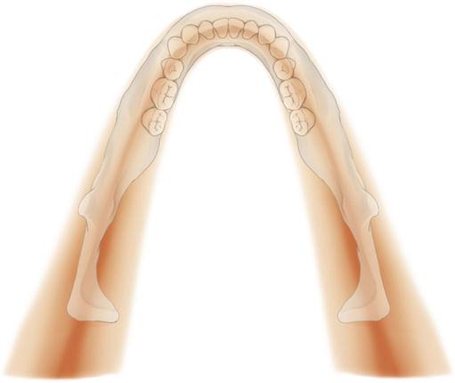
The focal trough is a three-dimensional curved zone, or “image layer,” where the structures lying within this zone are reasonably well defined on the final panoramic image (Fig. 10-7). The structures seen on a panoramic image are primarily those located within the focal trough. Images are most clear in the middle and become less clear further from the central line. Objects outside the focal trough are blurred, magnified, or reduced in size and are sometimes distorted to the extent of not being recognizable. The shape of the focal trough varies with the brand of equipment used as well as with the imaging protocol selected within each unit. The shape and width of the focal trough is determined by the path and velocity of the receptor and x-ray tube head, alignment of the x-ray beam, and collimator width. The location of the focal trough can change with extensive machine use, so recalibration may be necessary if consistently suboptimal images are being produced.
In some panoramic machines, the shape of the focal trough can be adjusted to conform better to the shape of the patients’ maxillomandibular anatomy or to show specific anatomic areas better, such as the TMJ or the maxillary sinuses. This adjustment is accomplished through varying the shape of the moving center of rotation and allows better imaging of children, unusually shaped patients, or specific anatomic sites of interest. For example, in some units, the rotational arc of the x-ray source-receptor movement is decreased to modify the focal trough size to pediatric jaws. The decreased rotational arc also results in reduced patient radiation exposure. In some panoramic units, the projection angle of the x-ray beam is modified to yield images with decreased overlap of adjacent teeth and with minimal superimposition of structures from the opposite side of the jaw.
Image Distortion
The panoramic image necessarily produces distortion of the size and shape of the object. These distortions make the panoramic image highly unreliable for linear or angular measurements. The image distortion is influenced by several factors, including x-ray beam angulation, x-ray source-to-object distance, path of rotational center, and position of the object within the focal trough. These parameters vary among panoramic units and among different regions of the jaws for the same unit. They are also strongly dependent on patient anatomy and positioning of the patient in the unit. These variables make it impossible to apply preset magnification factors that can be used to make reliable measurements on panoramic radiographs.
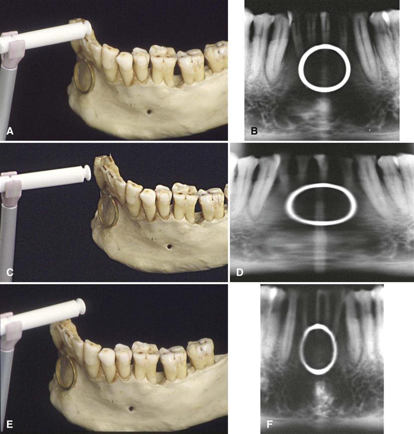
Horizontal magnification is determined by the position of the object within the focal trough. The magnitude of the horizontal distortion depends on the distance of the object from the center of the focal trough and thus is strongly influenced by patient positioning. Figure 10-8 illustrates the influence of patient positioning on image size and shape. Figure 10-8, A and B, shows a mandible supporting a brass ring properly aligned in the middle of the focal trough. Note the even magnification of the ring and the images of the anterior teeth in proper proportion. Figure 10-8, C and D, shows the same mandible positioned 5 mm posterior to the middle of the center of the focal trough. This position causes distortion of the ring in the horizontal dimension, with the ring appearing broader and a commensurate increased width of the images of the teeth. Figure 10-8, E and F, shows the same mandible positioned 5 mm anterior to the middle of the focal trough. The horizontal distortion results in the ring appearing narrow and a commensurate decreased width of the projected teeth. On these images, the vertical dimension, in contrast to the horizontal dimension, is little altered. These distortions result from the horizontal movements of the receptor and x-ray source. Thus, as a general rule, when the structure of interest, in this case the mandible, is displaced to the lingual side of its optimal position in the focal trough, toward the x-ray source, the beam passes more slowly through it than the speed at which the receptor moves. Consequently, the images of the structures in this region are elongated horizontally on the image, and they appear wider. Alternatively, when the mandible is displaced toward the buccal aspect of the focal trough, the beam passes at a rate faster than normal through the structures. In the example shown, because the receptor is moving at the proper rate, the representations of the anterior teeth are compressed horizontally on the image, and they appear thinner.
The same principle applies to the patient’s sagittal plane being rotated in the focal trough. The posterior structures on the side to which the patient’s head is rotated are magnified in the horizontal dimension because the posterior structures are moved away from the image receptor, whereas posterior structures on the opposite side are moved closer to the image receptor and are reduced in horizontal dimension. The resulting image has horizontally large molar teeth and mandibular ramus and severe premolar overlap on one side and horizontally smaller molar teeth and mandibular ramus on the other side. This imaging appearance must not be confused with a congenital or developmental facial asymmetry (this positioning artifact is demonstrated in Fig. 10-9).
Stay updated, free dental videos. Join our Telegram channel

VIDEdental - Online dental courses


