4 Forensics, Comparative Anatomy, Geometries, and Form and Function
Forensic Dentistry
Forensic dentistry (odontology) is the field within the greater disciplines of dentistry and forensic science that evaluates, manages, and presents dental evidence in legal proceedings in the interest of justice.1 Forensic dental casework often involves identification of unidentified or missing individuals, human remains, or victims of mass fatality incidents, including natural and accidental disasters. This is accomplished by comparison of a victim’s dentition and supporting structures with dental records of known individuals. The latter records may be obtained from private dental offices, prison or military dental databases, or records retained by the Federal Bureau of Investigation through its National Crime Information Center’s Missing, Unidentified, and Wanted Persons files in a Web environment.2
JAWS AND TEETH
Historically, despite this potential for an ever-changing dentition, inspection of the teeth and jaws has been used as a legally accepted method of human identification. Significant cases in which identification has been resolved by means of examination of dental evidence include identification of Revolutionary War hero Dr. Joseph Warren, the prominent Boston murder victim Dr. George Parkman, and Adolph Hitler.3
CHRONOLOGICAL AGE
The determination of dental maturity and tooth development has been considered in Chapter 2. As indicated there, the determination of chronological age is possible with some reasonable degree of accuracy. When it is employed in conjunction with evaluation of the stage of development of osseous tissues,4 the two methods can be used to determine an estimated age for an individual less than 20 years old. This is accomplished by entering data from both methods into an electronic encyclopedia on maxillofacial, dental, and skeletal development.5
Chronological age determination is a central issue in population studies, and racial and sex differences in tooth development and eruption patterns are acknowledged.6,7 Dental age assessment is also important in the forensic dental evaluation of human remains or living individuals. Aging of the dentition has been employed in forensic dental casework involving estimation of age of unidentified individuals.8,9
Separation of victims of multiple fatality incidents by age facilitates the narrowing of searches for eventual identification by comparison of medical and dental records. Dental evaluation of illegal immigrants, who may present authorities with misinformation concerning their age, is important in cases in which protection of unaccompanied minors is a concern.2
A variety of dental age estimation procedures have been employed. Some are useful in the analysis of radiographic changes associated with development of the dentition of children.10 Others use dental radiographs to determine the dental chronological age in living or deceased adult individuals.11,12
Estimation of the age of an adult may also be based on six characteristics first identified by Gustafson in 195013 by his observation of ground sections of extracted teeth. These six variables include attrition of the occlusal or incisal surfaces, degree of deposition of secondary dentin (evaluation of the size of the pulp chamber and canal), deposition of apical cementum, attachment level of the periodontium, root resorption, and radicular translucency.
In addition, biochemical methods that assess the ratios of levorotatory and dextrorotatory isomers of the long-lived, metabolically stable amino acid aspartate have been employed in both living and deceased subjects to age tissues in which this substance is found. Through a racemization process the L form of aspartic acid is slowly transformed into its stereoisomer, which is the D form of the amino acid. As enamel and dentin age, levels of the D form of aspartic acid increase in these calcified dental structures. These values can be measured and related to known levels for age estimation.14
DENTAL DNA
The most widely used method for forensic analysis of DNA material is the restriction fragment length polymorphism (RFLP) technique. This laboratory procedure requires that large amounts (>100 ng) of DNA be used in the analysis. The polymerase chain reaction (PCR) technique is employed when this cannot be accomplished because of degradation of the DNA molecule submitted as evidence, when only small amounts (<100 pg) of DNA are available for analysis, or when the DNA sample is fragmented.15 The PCR method amplifies the amount of DNA available for analysis by copying a specific locus of genetic material.16 Since only small amounts of DNA evidence need be evaluated using PCR technology, a positive identification may still be effected when human remains have been left unburied for long periods or have been incinerated, or when DNA trace evidence is obtained from saliva, blood, or fluid samples.
The calcified and pulp tissues within a tooth often present the forensic scientist with the most uncontaminated and protected DNA samples for analysis. Thus, even small amounts of DNA recovered from these tissues often may be analyzed using the PCR method when other means of identification have been lost or degraded as evidence.17,18
In addition, the sex of a decedent can be determined through DNA analysis of the sex-linked amelogenin gene. A DNA sample from the victim can be compared directly with one obtained from the clothing or other personal effects of a putative subject of interest or indirectly with a sample from the parents or siblings of the individual one is attempting to identify.19
BITE MARKS
When the dentition of a human or animal impresses the surface of an object during the act of biting, a pattern mark is often imprinted on that surface (Figure 4-1, A). Like a tool mark left as forensic evidence, the pattern left by the teeth can be evaluated and compared to the dentition that allegedly caused it (Figure 4-1, B). Whether the bite mark pattern involves the skin of a victim or suspect or remains on the surface of an inanimate object, to be probative, it must have class and individual characteristics consistent with a mark caused by teeth.
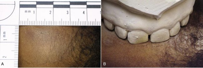
Figure 4-1 Forensic bite marks. A, Forehead bite marks. B, Bite marks related to maxillary dental cast.
(Courtesy Dr. David Ord.)
It is generally accepted that no two individuals have an identical dentition based on variations in the arrangement, spacing, size, and shape of specific teeth and dental arches. Currently, however, neither quantitative values nor databases have been established for the dentition that are similar to those described for fingerprint and DNA analysis and comparison. Thus, although bite mark evidence has been admissible in the federal and state courts of the United States based on the Frye rule and decisions related to the Federal Rules of Evidence,20,21 this evidence is often used in an exculpatory manner.
Guidelines have been established and are continually reviewed and revised to provide the forensic odontologist with evidence gathering and analytical procedures to be followed to ensure that recovered bite mark evidence is admissible in court and supports corroborative evidence in the case.22
Comparative Dental Anatomy
Figure 4-2 graphically illustrates a theory of the following four phylogenetic classes of tooth forms23–25:
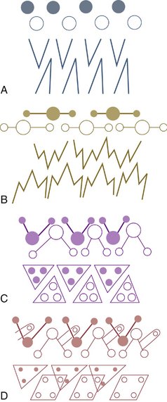
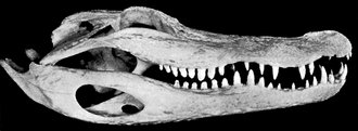
The haplodont class is represented schematically by the simplest form of tooth, the single cone (Figure 4-2, A). This type of dentition usually includes many teeth in both jaws and is seen where jaw movements are limited to simple open and close (hinge) movements (Figure 4-3). No occlusion of the teeth occurs in this class, since the teeth are used mainly for prehension or combat.26,27 Their main function is the procurement of food. Jaw movements are related to and governed by tooth form in all cases.
The triconodont class exhibits three cusps in line in posterior teeth as indicated in Figure 4-2, B. Anthropologically, the largest cusp is centered with a smaller cusp located anteriorly and another posteriorly. Purely triconodont dentitions are not seen, although certain breeds of dogs28 and other carnivores have teeth reflecting the triconodont form (Figure 4-4). Nevertheless, dogs and other animals carnivorous by nature are considered to be in the third category (Figures 4-5 through 4-8), namely, the tritubercular class.29,30 The three-cusp arrangement of the triconodont class and the more efficient three-cornered tritubercular molar arrangement are both consistent with the teeth’s bypassing each other more or less when the jaw is opened or closed. However, the quadritubercular class reflects an occlusal contact relationship between the teeth of the upper and lower jaws. Articulation of the jaws and teeth is consistent with jaw movements and functions of these classes of teeth.
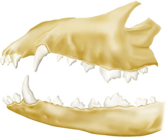
Figure 4-4 Permanent dentition of Canis familiaris.
(Modified from Tims HWM: Notes on the dentition of the dog, Anat Anz Bd 11:537, 1896.)
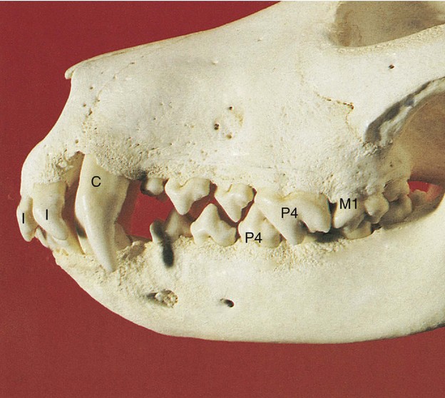
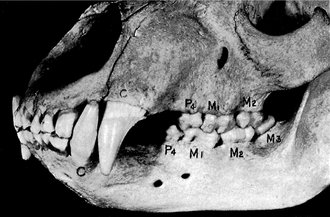
Figure 4-8 The bear. The extent of development of teeth and jaws and occlusion is often used to rate animal form. Compare with Figure 4-9. C, Canine; P, premolar; M, molar.
(From Kronfeld R: Dental histology and comparative dental anatomy, Philadelphia, 1937, Lea & Febiger.)
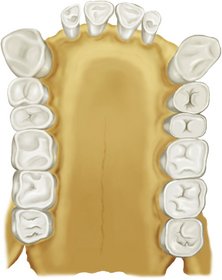
Animals that have dentitions similar to that of humans are anthropoid apes. This group of animals includes the chimpanzee, gibbon, gorilla, and orangutan (Figures 4-9 and 4-10). The shapes of individual teeth in these animals are amazingly close to their counterparts in the human mouth. Nevertheless, the development of the canines, the arch form, and the jaw development are quite different.
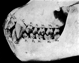
Figure 4-9 The ape. I, Incisor; C, canine; P, premolar; M, molar.
(From Kronfeld R: Dental histology and comparative dental anatomy, Philadelphia, 1937, Lea & Febiger.)
Stay updated, free dental videos. Join our Telegram channel
VIDEdental - Online dental courses


