23: Chronic Periodontitis

Chronic Periodontitis

H. Dommisch and M. Kebschull

Chronic periodontitis is the most prevalent form of periodontitis, and it generally demonstrates the characteristics of a slowly progressing inflammatory disease. However, systemic and environmental factors (e.g., diabetes mellitus, smoking) may modify the host’s immune response to the dental biofilm so that periodontal destruction becomes more progressive. Although chronic periodontitis is most frequently observed in adults, it can occur in children and adolescents in response to chronic plaque and calculus accumulation.

Chronic periodontitis has been defined as “an infectious disease resulting in inflammation within the supporting tissues of the teeth, progressive attachment loss, and bone loss.”18 This definition outlines the major clinical and etiologic characteristics of the disease: (1) microbial biofilm formation (dental plaque); (2) periodontal inflammation (e.g., gingival swelling, bleeding on probing); and (3) attachment as well as alveolar bone loss.

In addition to the local immune response caused by the dental biofilm, periodontitis may also be associated with a number of systemic disorders and defined syndromes. In most cases, patients with systemic diseases that lead to impaired host immunity may also show periodontal destruction. Therefore, periodontitis is a disease that is not only limited to the area of the oral cavity; it is also associated with severe systemic diseases (e.g., cardiovascular disorders, diabetes mellitus).40

This chapter discusses clinical features that have been described for chronic periodontitis. In addition, the disease’s etiology is summarized with the use of categories that explain the known factors (i.e., microbiologic, immunologic, and genetic) involved in the pathology of chronic periodontitis.

Clinical Features

General Characteristics

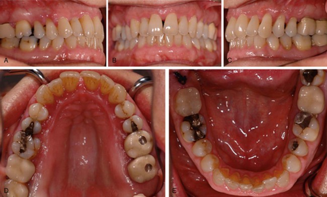
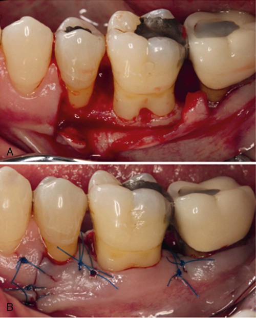
Characteristic clinical findings in patients with untreated chronic periodontitis include the following (see also the case presentation in Figures 23-1 through 23-6):

image
Figure 23-2 Documentation of the periodontal attachment level in the same patient as shown in Figure 23-1 at the time of the first visit. The red line displays the gingival margin as it reflects the recessions. Clinical attachment loss is illustrated by the filled (blue) area on the root surfaces. The deepest periodontal pocket was measured at 9 mm. Class I (green) and Class II (yellow) furcation involvements were documented. Bleeding with periodontal probing (i.e., gingival inflammation) is reflected by red dots. As a result of the patient’s history of smoking, the bleeding on probing score was relatively low, although the patient presented advanced attachment loss. Tooth mobility is indicated by the green line (tooth #19). (Reprinted from Kebschull and Dommisch, Thieme, 2012.)
image
Figure 23-3 Collage of a total of 11 radiographs showing the radiographic periodontal status at the time of diagnosis. Compare this with Figures 23-1 and 23-2. Note the generalized horizontal and localized angular and vertical bone loss on the mesial and distal sites of the molars. The radiographs show deep subgingival restorations (teeth #2 and #19), overhanging margins of restorations (teeth #14 and #15), a carious lesion (tooth #14), and insufficient root canal treatment (tooth #18). (Reprinted from Kebschull and Dommisch, Thieme, 2012.)
image
Figure 23-4 After the anti-infective therapy and periodontal reevaluation, resective periodontal surgery was performed for the patient introduced in Figures 23-1, 23-2, and 23-3. The surgical method involved an apically repositioned flap. A, Intrasulcular incisions at the buccal sites. Notice the Class I furcation involvement on tooth #14. B, Paramarginal incision at the palatal sites, with the excision of a distal wedge. C and D, Suturing with the use of 5-0 Prolene; buccal and palatal views, respectively. E, Occlusal view after suturing. F, Occlusal view 1 week after surgery. Tooth #14 received endodontic therapy and crown restoration before periodontal surgery. (Reprinted from Kebschull and Dommisch, Thieme, 2012.)
image
Figure 23-5 After the anti-infective therapy and periodontal reevaluation, resective periodontal surgery was performed for the patient introduced in Figures 23-1 through 23-4. The surgical method involved an apically repositioned flap. A, Intrasulcular incisions at the buccal sites. Note the Class I furcation involvement on tooth #19 and the horizontal bone loss affecting teeth #18, #19, and #20. B, Suturing with the use of 5-0 Prolene; buccal view. (Reprinted from Kebschull and Dommisch, Thieme, 2012.)
image
Figure 23-6 Documentation of the periodontal attachment level in the same patient introduced in Figures 23-1 through 23-5 after active periodontal therapy was completed and the supportive periodontal therapy was initiated. The red line displays the gingival margin as it reflects recessions after therapy. Clinical attachment loss is illustrated by the filled (blue) area on the root surfaces. The deepest periodontal pocket was measured at 4 mm. (Reprinted from Kebschull and Dommisch, Thieme, 2012.)

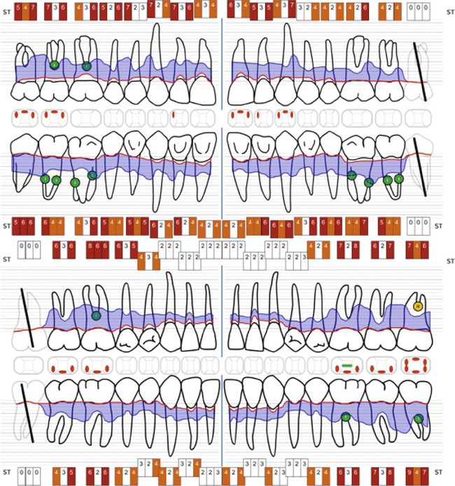
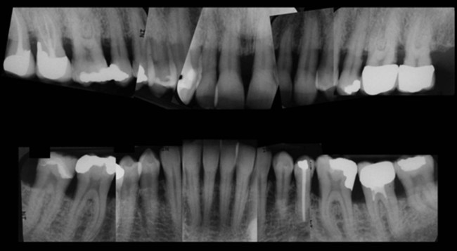
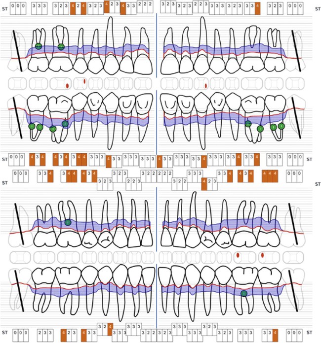
Chronic periodontitis can be clinically revealed with periodontal screening and recording, which results in a periodontal screening index rating. The condition is diagnosed via the assessment of the clinical attachment level and the detection of inflammatory changes in the marginal gingiva (see Figure 23-1). Measurements of periodontal pocket depth in combination with the location of the marginal gingiva allow for conclusions to be drawn regarding the loss of clinical attachment (see Figures 23-2 and 23-6). Dental radiographs display the extent of bone loss, which is indicated by the distance between the cementoenamel junction and the alveolar bone crest (see Figure 23-3). The distinction between aggressive and chronic periodontitis is sometimes difficult, because the clinical features may be similar at the time of the first examination. At later time points during treatment, aggressive and chronic periodontitis may be differentiated by the rate of disease progression over time, the familial nature of aggressive disease, the disease’s resistance to periodontal anti-infective therapy, and the presence of local factors.

Disease Distribution

Chronic periodontitis is considered a site-specific disease. Local inflammation, pocket formation, attachment loss, and bone loss are the consequences of direct exposure to the subgingival plaque (biofilm). As a result of this local effect, pocket formation and attachment as well as bone loss may occur on one surface of a tooth, whereas other surfaces maintain normal attachment levels. As a result of the site-specific nature, the number of teeth with clinical attachment loss classifies chronic periodontitis into the following types:

During chronic periodontitis, the local inflammatory response may lead to different patterns of bone loss, including vertical (angular) and horizontal bone destruction. Although vertical bone loss is associated with intrabony pocket formation, horizontal bone loss is usually associated with suprabony (supra-alveolar) pockets.

Symptoms

Chronic periodontitis is commonly a slowly progressive disease that does not cause the affected individual to feel pain. Therefore, most patients are unaware that they have developed a chronic disease that is also associated with other systemic diseases (e.g., cardiovascular disease). For the majority of the patients, gingival bleeding during oral hygiene procedures or eating may be the first self-reported sign of disease occurrence. As a result of gingival recession, patients may notice black triangles between the teeth or tooth sensibility in response to temperature changes (i.e., cold and heat). In patients with advanced attachment and bone loss, tooth mobility, tooth movement, and, in rare occasions, tooth loss may be reported. In those individuals with advanced disease progression, areas of localized dull pain or pain sensations that radiate to other areas of the mouth or head may occur. The presence of areas of food impaction may add to the patient’s discomfort. Gingival tenderness or “itchiness” may also be found.

Disease Progression

Patients appear to have the same susceptibility to plaque-induced chronic periodontitis throughout their lives. The rate of disease progression is usually slow, but it may be modified by systemic, environmental, and behavioral factors. The onset of chronic periodontitis can occur at any time, and the first signs may be detected during adolescence in the presence of chronic plaque and calculus accumulation. Because of its slow rate of progression, however, chronic periodontitis usually becomes clinically significant when a patient reaches his or her mid-30s or later.

Chronic periodontitis does not progress at an equal rate in all affected sites throughout the mouth. Some involved areas may remain static for long periods,33 whereas others may progress more rapidly. More rapidly progressive lesions occur most frequently in interproximal areas,32,34 and they may also be associated with areas of greater plaque accumulation and inaccessibility to plaque-control measures (e.g., furcation areas, overhanging margins of restorations, sites of malposed teeth, areas of food impaction).

Further factors that influence disease progression may be of systemic origin. Patients with poorly adjusted diabetes mellitus show a significantly higher risk of developing a severe progression of chronic periodontitis.43,61

Several models have been proposed to describe the rate of disease progression.57 In these models, progression is measured by determining the amount of attachment loss during a given period as follows:

• The continuous model suggests that disease progression is slow and continuous, with affected sites showing a constantly progressive rate of destruction throughout the duration of the disease.

• The random or episodic-burst model proposes that periodontal disease progresses by short bursts of destruction followed by periods of no destruction. This pattern of disease is random with respect to the tooth sites affected and the chronology of the disease process.

• The asynchronous, multiple-burst model of disease progression suggests that periodontal destruction occurs around affected teeth during defined periods of life and that these bursts of activity are interspersed with periods of inactivity or remission. The chronology of these bursts of disease is asynchronous for individual teeth or groups of teeth.

Stay updated, free dental videos. Join our Telegram channel

Jan 15, 2015 | Posted by in Periodontics | Comments Off on 23: Chronic Periodontitis

VIDEdental - Online dental courses

Get VIDEdental app for watching clinical videos