Dental hard tissue injury
Enamel infraction
Enamel fracture
Enamel-dentine fracture (uncomplicated crown fracture)
Enamel-dentine-pulp fracture (complicated crown fracture)
Crown-root fracture without pulp exposure
Crown-root fracture with pulp exposure
Root fracture
Alveolar fracture
Periodontal tissue injury
Concussion
Subluxation
Extrusion luxation (partial avulsion)
Lateral luxation
Intrusion luxation (central dislocation)
Avulsion
The most commonly observed injury to the dental hard tissues involves uncomplicated crown fractures of the maxillary anterior teeth [4]. Crown fractures account for up to 76 % of the dental trauma reported to the permanent dentition. This consists of enamel infraction, enamel fracture, uncomplicated crown fracture, complicated crown fracture and crown-root fractures [4].
Of the periodontal tissue injuries, luxation injuries were consistently found to be the most frequent dental trauma involving the periodontal tissue in studies followed by subluxation. Almost one-third (31.6 %) of teeth suffered a combination of tooth fracture and luxation injury. Furthermore, two-thirds of all combination injuries were observed amongst teeth with minor luxation injuries such as concussion and subluxation [5–8].
The avulsion of permanent teeth is considered to be one of the most serious dental injuries to occur and is seen in 0.5–3 % of all dental traumas. In the worst case scenario the tooth is lost or extracted due to the complications following replantation. The loss of an upper central incisor has a significant effect on dental and facial aesthetics due to the prominence of the maxillary teeth, and this can affect an individual’s self-esteem and general social interaction [9, 10].
Infraction injuries are microcracks in the enamel (incomplete enamel fracture without loss of tooth structure). They can be relatively common as a direct result of impact trauma to a tooth requiring no immediate treatment. Pulpal complications are considered to be rare. Pulpal necrosis has been reported in 1.6 % of teeth with enamel infraction and concomitant enamel fracture, whilst 2.3 % of teeth with enamel infraction and concomitant enamel-dentine fracture developed pulpal necrosis. Interestingly, when enamel infraction occurred in isolation, 3.5 % developed necrosis. This has been attributed to the traumatic impact being dissipated through the pulp rather than transfer of energy directly to the supporting tissues resulting in loss of tooth structure. The rate of pulpal necrosis for enamel infraction with a concomitant injury from a concussion or luxation injuries has been shown to range from 14.7 to 60.0 %. Furthermore, the state of root development has been found to be associated with the risk of developing pulpal necrosis (more common in a mature fully developed closed apex teeth). Pulp treatment is rarely indicated and only carried out following definitive signs and symptoms of irreversible pulpitis or pulpal necrosis with infection. The clinician should bear in mind that reactions to pulp sensibility testing in traumatised teeth are lowered immediately following trauma requiring longer observation times before a definitive decision can be made regarding the likely status of the pulp. As with all traumatic injuries, further follow-up after the initial injury is advised [1, 4, 11–14].
Enamel fractures without the exposure of the dentine result in the loss of the enamel structure. The mesial or distal corners of the maxillary central incisor teeth usually sustain this type of injury. Since loss of tooth structure is confined to enamel, patients are asymptomatic not requiring emergency treatment. Pulp survival following enamel fractures has been found to be high (>99 %). However, when there is a concomitant luxation injury, the chances of pulp survival can decrease to 33.3 %. The stage of root development is associated with the potential of pulpal healing following enamel fractures. The risk of pulpal necrosis in teeth that suffered enamel fracture with incomplete root development and open apices was 1.0 % compared to the risk of 2.5 % in teeth with complete root development and closed apices [5–8, 11–15].
When an enamel-dentine fracture without pulpal involvement occurs, the treatment objective is to maintain pulp vitality and restore aesthetics and function. Direct dentine exposure can result in discomfort from temperature changes. Additionally, the enamel-dentine fracture may result in a near pulpal exposure. Emergency treatment prior to definitive restoration includes protection of the exposed dentine. When the fractured crown is retained, bonding and reattachment is considered more aesthetically desirable than the use of tooth-coloured restorative materials. The prognosis of pulpal healing has been reported to range from 94.0 to 100 %. Both extent of dentine exposure and presence of a concomitant injury affecting the periodontal tissues can have an adverse effect on pulp survival. In enamel-dentine fractured teeth with a concomitant luxation injury, 16.8–69.9 % have been reported to develop pulpal necrosis [1, 11–16].
The treatment of the enamel-dentine fracture involving the pulp (complicated crown fracture) is aimed at removing the possibility of bacterial ingress into the pulp, thereby allowing the pulp time to recover and repair. Various treatment modalities including direct pulp capping, partial pulpotomy and pulpectomy have been recommended depending on extent of pulpal exposure and the time elapsed between injury and treatment provided.
In young patients with immature (developing) teeth, it is advantageous to try and maintain pulp vitality by either pulp capping or partial pulpotomy procedures. Pulp capping is indicated when a small exposure is present and can be treated shortly after the dental trauma occurs. When the pulp is more severely exposed and there has been a time delay, then the extent of pulpal inflammation may result in irreversible damage of the superficial layer of the pulp. If an enamel-dentine-pulp fracture occurred, the removal of a minimum of 2 mm of pulp beneath the exposure is required to remove the inflamed pulp. This has given rise to the procedure known as ‘partial pulpotomy’. Alternative levels of pulpal amputation are recommended based on the extent of pulpal inflammation as determined clinically by the presence of pulpal haemorrhage following a dental trauma. In instances where pulpotomy cannot be performed due to the magnitude of pulpal inflammation, pulpectomy is indicated. Following pulpal therapy, the fractured tooth can be restored in a similar manner to an enamel-dentine fracture. The long-term prognosis of the enamel-dentine-pulp-fractured tooth depends on the presence of other concomitant injuries, time elapsed between pulp exposure and treatment, bacterial infection and the stage of root development. Long-term studies of enamel-dentine-pulp fractures treated with partial pulpotomy and using calcium hydroxide showed a success rate of 87.5 % after a follow-up period of 7.5–11 years. The teeth that did not recover required endodontic treatment due to a new injury or for aesthetic purposes [1, 14, 17–20].
Crown-root fractures involve the enamel, dentine and part of the root (cementum) surface of the tooth. The fracture line invariably passes subgingivally often exposing the pulp requiring endodontic therapy if the tooth is to be retained. The treatment of a crown-root fracture is challenging and further complicated by the extent of the subgingival fracture often requiring a multidisciplinary approach. Treatment options include periodontal surgery to expose crown margins, restorative management only with extension of the margins of the restoration below the level of the gingival margin, orthodontic extrusion, intentional replantation (surgical repositioning), autotransplantation, root submergence (decoronation), extraction and replacement or orthodontic space closure. Treatment of crown-root fractures can be complex and time consuming, but often teeth with these types of fractures can be saved. In an adult patient, implant replacement is sometimes a viable alternative. In the case of a growing patient with a tooth that is not restorable, root submergence (decoronation) may be indicated to preserve the bone and allow for normal alveolar development prior to implant placement when growth is complete [21–28].
Root fractures are relatively uncommon, making up 0.5–7.7 % of dental trauma. The maxillary permanent central incisors are most frequently affected (up to 80 % of cases). They involve the cementum, dentine and the pulp and can present with or without clinical signs of luxation of the coronal fragment. The fracture can appear radiographically as a single line or multiple lines across the root in the apical, middle or coronal 1/3. Radiographic diagnosis of root fractures requires multiple views at different vertical angles to obtain a three-dimensional image. Root fractures can occur in either the cervical, middle or apical portion of the tooth.
Many root fractures heal without intervention in one of three modalities: hard tissue interposition, interposition of the bone and periodontal ligament or interposition of the periodontal ligament alone. In the past, rigid splints were used for an extended period of time (up to 4 months); however, no additional benefits were found with regard to pulpal or periodontal healing. The prognosis of root-fractured teeth appears to be multifactorial related to the position of the root fracture, the stage of root development, presence of bacterial infection and the type of healing pattern that occurs. Pulpal necrosis of the coronal fragment has been reported in up to 25 % of teeth with root fractures. Pulp canal obliteration (partial or complete) may occur in up to 70 % of cases. Obliteration of the apical root canal is commonly seen in cases of calcified tissue healing. When interposition of either the connective tissue or the connective tissue and bone occurs between the fragments, obliteration occurs more likely in both apical and coronal fragments. Interestingly, treatment delays showed no significant effect on the frequency and type of healing after accounting for factors such as root development and the extent of displacement. Root-fractured teeth should be splinted for up to 4 weeks initially and then reviewed. In cases where the root fracture has occurred, coronally extended splinting times may be necessary to ensure adequate stabilisation and healing occurs. Follow-up will be required to monitor pulpal status and fracture healing [29–46].
Jaw fractures can involve the base of the mandible or maxilla and often the alveolar process. Tooth involvement is common in mandibular and maxillary fractures and may require additional endodontic management. Nonsurgical treatment of bone fractures involves immobilisation, which for the facial bones is achieved with either maxillomandibular fixation (MMF) using dental fixed arch bars or direct internal fixation using screws and plates. Associated tooth injuries are difficult or impossible to treat during immobilisation, so that some treatment is necessary beforehand (e.g. coverage of exposed dental areas, temporary filling of crown fractures, repositioning of luxated teeth and endodontic treatment if the pulpal vascular supply is lost in the accident). Teeth in the line of a mandibular fracture should not be extracted as a first-aid measure unless they impair repositioning of the jaw fragments [47–54].
Luxation injuries represent the majority of dental trauma in the primary teeth and are the second most common type of injury sustained in the permanent dentition. The extent of injuries to the periodontal tissues is largely determined by the resilience of the underlying periodontal structures. Periodontal injuries often result in displacement of the affected tooth. Treatment of the displaced tooth aims to reduce the amount of dislocation from the tooth socket by repositioning. Common endodontic complications include pulp necrosis with infection, pulp canal calcification, ankylosis and root resorption. Factors that affect the prognosis of luxated teeth include the degree of displacement, delayed treatment time, root maturation and concomitant crown fractures. Most cases of pulp necrosis in luxated teeth become evident with the first 4 months. Root resorption often occurs within the first 6 months after injury and can develop quite rapidly, particularly in immature teeth. Hence, frequent follow-up examination is recommended. In some cases, pulp necrosis may appear at a much later date, and therefore long-term follow-up is also essential.
Concussion and subluxation injuries result from disturbances and/or laceration of the periodontal ligament fibres with very little disturbance to the neurovascular supply. These teeth display little displacement from the bony socket and therefore repositioning and splinting is seldom prescribed. Marked tenderness to percussion but no abnormal loosening or displacement of the traumatised tooth characterises a concussion injury to the supporting structures. Reported frequency of pulpal necrosis ranges from 3 %, and 2–7 % may undergo further pulp canal calcifications. Subluxation injuries are characterised by abnormal loosening of the tooth but without any displacement. Teeth are tender to percussion, and there may be some bleeding in the gingival crevice. Prognosis for subluxation injuries is generally good. Reported frequency of pulp necrosis ranges from 6 to 26 %. Pulp canal calcification has been reported to occur in 9–12 % of cases and progressive root resorption in less than 2 % [1, 6, 55–59].
Extrusive luxation of the tooth presents as a displaced tooth extruded from its socket. Emergency treatment requires repositioning and splinting. A delay in treatment usually results in difficulty in correct repositioning due to an established blood clot. In teeth that suffered an extrusive luxation injury, 26–80 % developed pulpal necrosis. The stage of root development significantly influenced the risk of pulpal necrosis development, with immature roots having a risk of 5.9 % compared with 56.5 % for teeth with complete root development. Root resorption, mainly in the form of inflammatory or surface resorption, was consistently observed in 5.5–9.4 % of extruded teeth [1, 6–8, 60–64, 91].
A lateral luxation injury is associated with a comminution or fracture of the alveolar socket. The tooth may be immobile as the injury crushes the bony socket walls. Emergency treatment for a laterally luxated tooth involves separation of the displaced tooth from the bony lock, repositioning the tooth and then stabilisation for at least 4 weeks. In laterally luxated teeth, the risk of developing pulpal necrosis was found to be between 33 % and 58 % and is related to the stage of root development. When the roots are immature, the risk of developing pulpal necrosis is reported to be 4.7 % compared to the risk of 65.1 % for mature roots. External root resorption is reported in 27 % of laterally luxated teeth and is predominantly surface resorption. The risk of developing inflammatory resorption is relatively low (0.8 %) [1, 5, 7, 8, 63–66, 91].
Intrusive luxation injuries result in a tooth being displaced in an axial position into the alveolar bone. This type of injury results in compression of the periodontal ligament and is accompanied by comminution or fracture of the alveolar socket. The emergency management of an intruded tooth may involve passive repositioning with spontaneous re-eruption or active repositioning with orthodontically guided eruption or surgical repositioning. For intruded teeth with immature root development, spontaneous eruption is preferred. For patients above 17 years of age, active repositioning with surgical or orthodontic intervention is recommended. The type of repositioning depends on the stage of root development and also on the extent of intrusion. For teeth with up to 3 mm of intrusion, passive repositioning is recommended; for 3–7 mm intrusion cases, orthodontic repositioning is preferred; for more than 7 mm of intrusion, surgical intervention is recommended. Of all the periodontal injuries, intrusive luxations are considered to be the most severe due to both the disruption of the vascular supply and the damage to the periodontal ligament cells. Pulpal necrosis is observed in 44–96 % of intruded teeth. Additionally, root resorption is found in up to 80 % of intruded teeth, of which 40 % resulted in replacement resorption [67–73, 91].
Avulsion or exarticulation occurs when a traumatic injury totally displaces a tooth from the socket resulting in potential for periodontal ligament damage and alveolus fracture. Although the prognosis for an avulsed tooth must always be guarded, replantation as soon as possible followed by a brief period of flexible splinting and endodontic therapy has been shown to be the most effective method of treatment. The shortest extra-oral period (less than 15 min), the minimum manipulation of the tooth surface and the socket and the use of an appropriate storage medium (milk, saliva and Hanks’ balanced salt solution) have been identified as important factors that minimised subsequent root resorption.
The most common reason for unfavourable long-term survival of avulsed teeth is root resorption. A number of factors have been identified as being important in the prevention and management of root resorption associated with avulsed teeth including the stage of root development, the viability of the periodontal ligament cells and amount of damage sustained, the tooth storage conditions and the duration of time prior to replantation (extra-alveolar drying time).
The best outcome for a tooth avulsion is when the tooth can be replanted within a few minutes after the accident. The prognosis of a replanted avulsed tooth depends on the survival of both the periodontal ligament cells and the pulp. A large percentage of teeth that are replanted within 15 min will result in viable periodontal ligament cells and potential for reattachment within a few weeks. The extra-oral alveolar time has been shown to have detrimental effects on the survival of the periodontal ligament cells (approximately 20 min), and an inadequate storage medium often results in replacement resorption. If an avulsed tooth is left dry for more than 1 h, the odds of periodontal ligament cell survival are very poor. Despite the risk of ankylosis and replacement resorption, it may still be worthwhile replanting the tooth, particularly in a young patient, where several more years of use may be attained before eventual loss of the tooth [1, 10, 74–90].
Transient apical breakdown is a process that can develop as a result of certain traumatic injuries (subluxation, extrusion and lateral luxation) to teeth and their supporting tissues. Radiographically, a periapical radiolucency develops with the affected tooth that resolves without complication and does not require any endodontic intervention. The incidence of transient apical breakdown is relatively low and was reported to be 4.2 % of luxated cases. The condition was associated with either a pronounced radiolucency appearing spontaneously within a short time after injury or with a persistent expansion of the periodontal ligament space progressing over an extended period. During follow-up periods, the radicular and bony conditions either had returned to normal or showed evidence of surface resorption and/or root canal obliteration without further complications [60].
Pulp canal obliteration (calcification of the root canal) is a common sequel following luxation injuries to permanent teeth, particularly teeth that have been injured before their root formation has been completed. Clinically, a yellowish discolouration of the crown may be observed. Pulp canal calcification is also a common occurrence in root-fractured teeth occurring principally in the region of the fracture and in the apical fragment. It may also occur in teeth associated with alveolar and jaw fractures. In most traumatised teeth that have pulps undergoing calcification, the hard tissue is deposited longitudinally along the dentinal walls of the pulp canal. This gradually diminishes the pulp in size until it can barely be observed radiographically. Endodontic treatment is not indicated unless there is evidence of irreversible pulpitis or pulpal necrosis. Pulp necrosis and infection may occur up to 20 years following injury. Assessment of the status of the pulp is difficult since these teeth do not usually respond to thermal pulp sensibility testing. Most, however, do respond to electrical stimulus, and therefore electric pulp sensibility testing is the desirable method for assessing the status of the pulp in calcified teeth [92–94].
Root resorption is a common sequel to dental trauma and may be caused either directly by the traumatic incident or indirectly through subsequent infection. The three most common types of resorption following trauma include surface resorption, replacement root resorption and inflammatory root resorption. Surface resorption is believed to be a self-limiting response to a localised injury to the periodontal ligament or cementum. In traumatised teeth, this type of resorption occurs more commonly in the apical portion of the root and may be seen as a shallow rounding of the external root shape. Replacement root resorption, generally associated with luxated or replanted avulsed teeth, results in the replacement of tooth structure by the bone and can be recognised radiographically by the diffuse nature of the resorption defect and the disappearance of the periodontal space adjacent to the area of the resorption. This progressive type of resorption has a very poor prognosis. Irreversible damage to the periodontal ligament leads to a fusion between the dentine and bone with progressive replacement of the dentine by the bone.
Inflammatory root resorption occurs as a result of a necrotic and infected pulp, which can be recognised radiographically by the development of radiolucency in the bone adjacent to the resorptive defect. Removal of the infected pulp tissue, dressing with an anticlastic medicament and subsequent filling of the root canal with gutta-percha will usually halt the resorptive process [92, 95, 96].
The management of dental traumatic injuries would be incomplete without a discussion of the need to fully assess a patient with dental injuries including dental hard and soft tissues and injuries to supporting tissues and to the patient as a whole. All traumatic dental injuries need to be followed up over time. Follow-up procedures include a clinical examination, a radiographic assessment and pulp sensibility testing [97]. Recommendations for follow-up examinations for injuries are in accord with those recommended by the International Association of Dental Traumatology [1, 14, 15, 20, 27, 44, 51, 58, 60, 63, 73, 90].
Routine radiographic examinations for preoperative, perioperative and postoperative monitoring of complications and healing of dental trauma cases should include a parallel periapical radiograph with the central beam projected through the tooth in question and a lateral angulation from either the mesial or distal aspect of the tooth. The latter allows enhanced visualisation of root fractures that may not be noted when the beam is aimed centrally [98, 99].
Splinting of teeth with luxation injuries, avulsions and root fractures is often necessary to stabilise a tooth in position and to assist in periodontal and pulpal healing. A non-rigid flexible splint has been shown to be the most desirable. An ideal splint should amongst other things be easily fabricated in the mouth without additional trauma; be passive, allow for physiological mobility; be non-irritating to the soft tissues, not interfere with the occlusion; allow endodontic access; and be easily cleaned and easily removed. Recommended splinting times are up to 2 weeks for most avulsion and luxation injuries unless they occur in association with alveolar fractures; up to 4 weeks for lateral luxation injuries, alveolar fractures and root fractures; and up to 4 months for cervical-third root fractures. Many dental injuries do not occur singly. Thus, these splinting times cannot be rigorously applied. In general, splinting times have to be adjusted to accommodate the more major injuries [100–102].
There is limited evidence for the adjunctive use of systemic antibiotics for luxation injuries and no current evidence that antibiotic coverage improves the outcome for root-fractured teeth. The use of intra-canal antibiotics and corticosteroids immediately after replantation appears to halt the progression of inflammatory root resorption, although replacement resorption still occurs to some extent. Antibiotic coverage may be warranted by the patient’s medical health and/or injuries sustained, and the decision to provide any coverage lies with the individual practitioner in line with current guidelines [54, 103, 104].
Pulp sensibility testing immediately after trauma is an unreliable means of predicting the true status of the pulp. A negative response is often a common finding immediately after the pulp has suffered trauma. Teeth that initially give a negative response may prove to respond positively in the coming months. Endodontic treatment is only indicated for those teeth in which pulp necrosis has occurred. At least two signs and symptoms (clinical and/or radiographic) are necessary to make the diagnosis of a necrotic pulp. Responses to sensibility testing need to be followed up over time before a definitive diagnosis of pulp necrosis is made [105, 106].
Treatment planning for the management of traumatic injuries should be based on sound biological principles that form the basis of current guidelines. Careful adherence to treatment protocols with these goals in mind should allow for optimal healing and retention of the tooth where possible [107].
15.2 Initial Management and First Aid
Initial management of any trauma should consider the general wellbeing of the patient and any loss of consciousness; acute bleeding, respiratory problems or neurological signs/symptoms may indicate life-threatening conditions requiring urgent medical attention. Often the patient presenting with dental trauma in your office will be stable, showing normalisation of vital functions. The secondary survey starts with the initial history taking and should include general patient details, general medical health of the patient, past tetanus status and questions related to the injury (when, where and how the injury occurred). It is highly pertinent that all information should be obtained in chronological order with contemporaneous notes recorded including time and date of the trauma. This is particularly relevant when an assault is alleged resulting in legal ramifications or insurance matters related to trauma at school or the workplace. It is important for the clinician to calm and reassure the often-distressed patient prior to any initial examination.
Extra-orally simple visual inspection can assess facial symmetry, swelling, facial muscle movements (cranial nerve VII) and mouth opening and deviation (including maximal inter-incisal distance). Clear draining fluid from either the nose or ears may indicate cerebrospinal fluid, which indicates severe cranial damage. Palpation noting any marked tenderness, bony steps, mobility/crepitus and facial sensation abnormalities (cranial nerve V) can give further clues to any underlying facial bone fractures. Numbness of the lower lip in the context of a facial trauma can be highly suggestive of a mandibular fracture. Numbness of the cheek, nose, upper lip or maxillary teeth may indicate a complex fracture involving the orbitozygomatic complex. Numbness of the maxillary teeth alone may indicate an orbital floor fracture affecting branches of the middle to anterior superior alveolar dental nerve.
Dental injuries can often result in facial or lip lacerations that may require suturing or referral to an appropriate specialist (Figs. 15.1 and 15.2). Before examination of the mouth, a thorough facial examination should have been carried out including assessment of frontal, naso-orbital, nasal, middle-third, zygomatic, orbital and mandibular fractures. Inability to open the mouth, limited opening or deviation may all indicate mandibular or temporomandibular joint fractures. Mandibular fractures will usually be associated with painful, limited opening, and if a condyle is involved, then the jaw may deviate towards the side of the fracture. A deranged occlusion may also indicate a fracture of the mandible or zygoma until proven otherwise. Appropriate referral to an oral maxillofacial surgeon may be required.
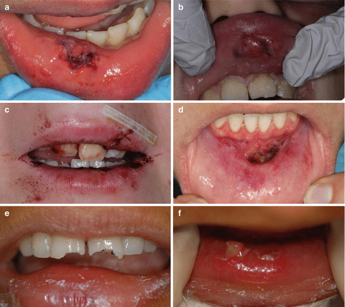
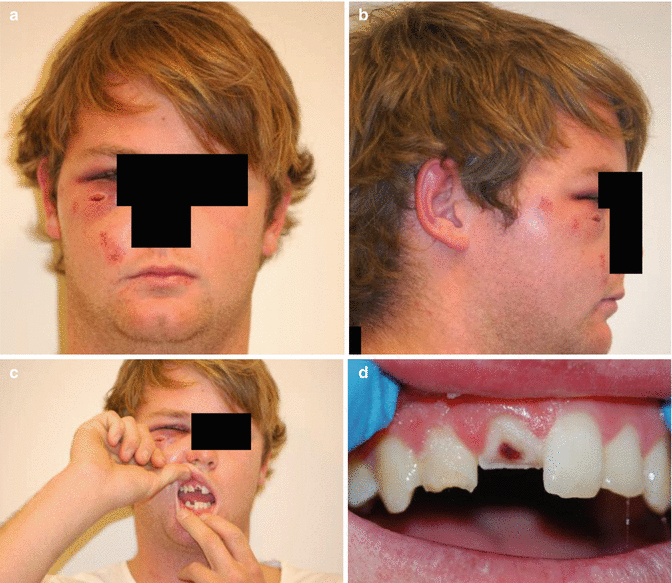
Fig. 15.1
Clinical photographs demonstrating soft tissue lacerations associated with traumatic injuries. Note: (a, b) lacerations affecting lips – simple resorbable sutures can be used for primary closure. Deep sutures may be required for muscle repair. (c) Laceration affecting the upper lip with vermillion border involvement. Correct suturing will be required to align vermillion border with appropriate skin sutures. (d) De-gloving injury affecting the alveolar mucosa. (e, f) Uncomplicated maxillary central incisor crown fractures embedded in the lower lip (Pictures courtesy of Drs K Dang, A Timmermann, and D Felman)
Fig. 15.2
Clinical photographs demonstrating soft tissue lacerations associated with traumatic injuries. Note: (a, b) facial laceration affecting right infraorbital area with swelling and bruising. (c, d) Intraoral injury. A complicated crown fracture affecting tooth 11; an uncomplicated crown fracture associated with tooth 12 (Pictures courtesy of Dr K Dang, A Timmermann, and D Felman)
Intra-oral examination should include assessment of the crowns of the teeth (checking for signs of possible fracture or pulp exposure), displaced teeth (luxation injuries) and missing teeth, occlusion disturbances and abnormal mobility of individual or groups of teeth and surrounding soft tissues (including degloving injuries). All missing teeth and fragments should be accounted for (Figs. 15.1 and 15.2).
Radiographic assessment will be invaluable in the diagnosis and management of dental trauma. Extra-oral plain film radiography or cone beam computer tomography is indicated when suspected facial fractures including jaw and condylar fractures are suspected. Soft tissue radiographic assessment is indicated when tooth fragments or foreign objects might have been embedded into the lips. Pretreatment radiographs are essential to determine the size of pulp chamber, the stage of root development and the appearance of the periodontal ligament and presence of any root fractures. Parallel periapical radiographs are also essential to monitor any changes in the pulp space, resorption and calcifications that may indicate future therapeutic intervention.
15.3 Crown Fractures
Enamel infraction
Clinical and radiographic findings
An incomplete fracture (crack) of the enamel has occurred without loss of tooth structure (Fig. 15.3). The use of indirect light or transillumination or dyes may help visualise the craze line. Clinical examination reveals no abnormal tenderness to percussion or palpation. Radiographic appearance reveals no abnormalities.
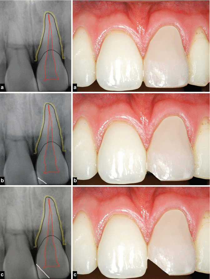
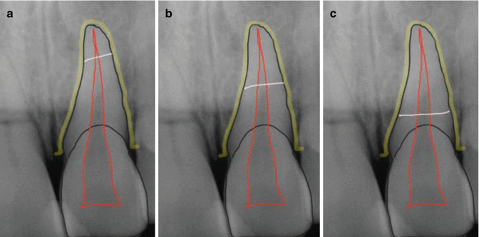
Fig. 15.3
Diagrams representing the different types of crown/root fractures that may present following a traumatic injury. (a) Enamel infraction requiring no treatment. (b) Uncomplicated crown fracture – enamel only. Reattachment of the tooth fragment or tooth colored restoration will protect the underlying pulp. (c) Uncomplicated crown fracture – enamel and dentine. Dentinal tubule exposure can result in sensitivity and pulpal inflammation if left untreated
Treatment objectives
Often no treatment is usually indicated. In cases of marked infraction, etching and sealing with a composite resin may prevent future discolouration of the fracture line.
General prognosis
Pulpal complications are rare.
Recommended follow-up
No further follow-up is generally needed.
Enamel fracture (uncomplicated)
Clinical and radiographic findings
Clinical findings reveal loss of tooth structure confined to enamel (Fig. 15.3). Clinical examination reveals the tooth exhibits no abnormal pain or tenderness. Normal mobility is noted and pulp sensibility testing is normal. Radiographic examination of the soft tissues (lips) may be recommended to rule out the presence of any tooth fragments or foreign material potentially displaced into the lips. Two periapical films should be taken with either a mesial or distal tube shift to rule out the presence of any underlying root fractures.
Treatment objectives
The aim of any proposed treatment is to maintain pulp vitality and restore aesthetics and function. Minor fractures or rough margins/edges should be smoothed. If the tooth fragment is available, then re-bonding may be possible using composite resin and the acid-etch technique. When unavailable, restoration with a composite resin restoration will achieve the desired aesthetic outcome.
General prognosis
Overall prognosis is good although any concomitant periodontal injury sustained can have an adverse affect on outcome.
Recommended follow-up
Due to the unpredictability of dental trauma, pulp vitality testing should be carried out both immediately after the occurrence of the injury and again in 6–8 weeks and 1 year.
Enamel-dentine fracture (uncomplicated)
Clinical and radiographic findings
Crown fractures involving enamel and dentine without pulpal exposure are called uncomplicated crown fractures (Figs. 15.3 and 15.5). Clinical examination reveals loss of tooth structure confined to enamel and dentine with potential exposure of dentinal tubules that can lead to pulpal inflammation. Radiographic examination confirms enamel-dentine loss. Soft tissue radiographic examination of the lips may be indicated to ensure no fragments are present. Presence of root fractures should also be assessed using two parallel periapical radiographs with mesial or distal cone shift technique.
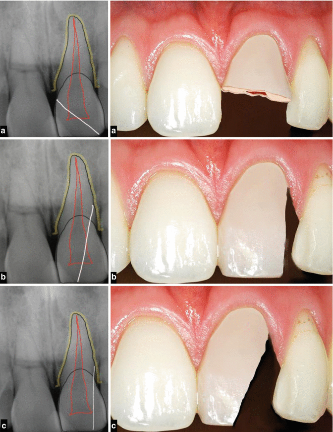
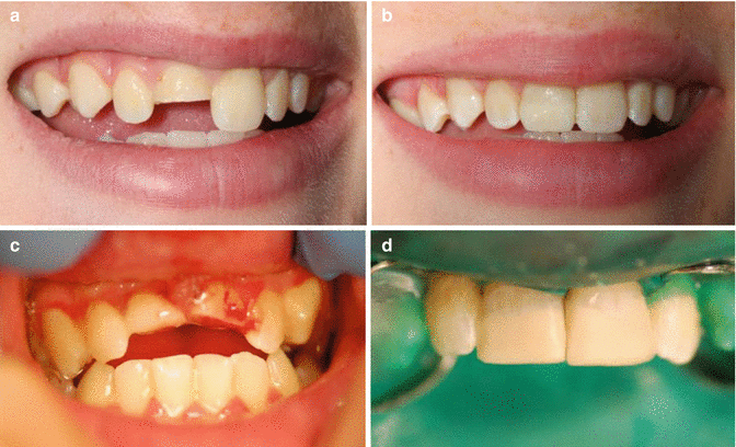
Fig. 15.4
Diagrams representing the different types of crown/root fractures that may present following a traumatic injury. (a) Complicated crown fracture – enamel, dentine, and pulp. Treatment options include either pulp preservation procedures (direct pulp capping or Cvek pulpotomy) or pulpectomy procedures. Pulp preservation should be attempted in immature teeth with incomplete root development. Conversely, in mature teeth with extensive loss of tooth structure, root canal therapy is sagacious before cast restoration. (b) Complicated crown-root fracture – enamel, dentine, and pulp. (c) Uncomplicated crown-root fracture – enamel and dentine only
Fig. 15.5
Clinical photographs demonstrating uncomplicated crown fractures. Note (a, b) uncomplicated crown fracture affecting tooth 11 restored with composite resin restoration. (c, d) Uncomplicated crown fractures affecting teeth 11 and 21 with appropriate restorations (Pictures courtesy of Drs K Dang and A Timmermann)
Treatment objectives
The aim is to preserve the vitality of the underlying pulp by sealing the exposed dentinal tubules and restore aesthetics and function for the patient. If the tooth fragment is available, it may be possible to reattach the fragment; otherwise, direct application of dentine bonding agents and the use of bonded tooth-coloured restorations will be effective. Reattachment of the tooth fragment requires isolation of the tooth under rubber dam. Both the tooth fragment and fractured tooth should be cleaned with pumice and water prior to reattachment. Accuracy of fit should be checked prior to the use of acid-etch technique according to manufacturer directions and type of bonding system used. Cases where exposed dentine encroaches to within 0.5 mm of the underlying pulp (determined by visible pink pulp tissue beneath the exposed dentine) will require application of an indirect pulp capping material.
General prognosis
The survival of the pulp and formation of tertiary dentine (reactionary) or pulpal necrosis depend on a number of factors including residual dentine thickness, age of the patient, concomitant periodontal injury and the length of time between trauma and treatment intervention. The overall prognosis for these types of fractures is generally good.
Recommended follow-up
Periodic evaluation is necessary to determine pulpal status and whether pulp necrosis has developed. The patient should be reviewed 6–8 weeks following treatment and at 1 year.
Crown fracture (complicated)
An enamel-dentine fracture with pulp exposure.
Clinical and radiographic findings
Crown fractures involving enamel, dentine and pulp are known as complicated crown fractures (Fig. 15.4 and 15.6). The degree of pulpal involvement can range from a pinpoint exposure to complete coronal pulp exposure. Clinically the exposed pulp may be sensitive to stimuli. Radiographic examination confirms pulpal involvement.
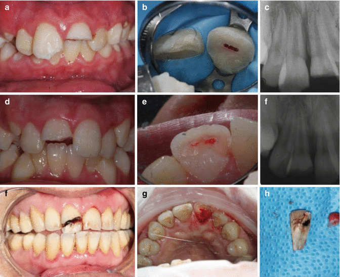
Fig. 15.6
Clinical photographs and radiographs demonstrating complicated crown fractures (enamel-dentine-pulp injuries). Note: (a–c) complicated crown fracture affecting tooth 21. (d–f) Complicated crown fracture affecting tooth 21. (f–h) Complicated crown fracture affecting tooth 21 (Pictures courtesy of Drs K Dang, A Timmermann, and D Felman)
Treatment objectives
Following pulp exposure, bacterial contamination precludes any healing and repair unless the exposure is treated and protected. The initial reaction to pulpal exposure is haemorrhage at the site followed by a superficial inflammatory response. The longer the pulp is exposed before adequate protection, the greater the risk of either destructive (necrosis) or proliferative changes (pulpal polyp). Treatment modalities include direct pulp capping procedures, partial pulpotomy (Cvek), and full pulpotomy or pulpectomy procedures. Vital pulp therapy procedures in young patients with immature developing, thin, weak roots offer the potential for continued root development. Conversely in the fully developed permanent dentition, pulpectomy procedures may offer a better chance of success rather than risking the development of apical periodontitis following direct pulp capping or pulpotomy procedures.
General prognosis
Overall prognosis of attempts to maintain pulp vitality as opposed to complete pulpectomy procedures is dependant on any underlying concomitant periodontal injury sustained, age of the pulp exposure and stage of root development (incomplete versus complete). Treatment modalities aimed at preserving pulp tissue may result in either failure in the future or degenerative pulp changes such as pulpal calcification.
Recommended follow-up
Periodic evaluation both clinically and radiographically should be carried out at 6–8 weeks and 1 year following treatment. Pulp preservation treatments should be assessed for continued pulp vitality ensuring necrosis has not developed.
Crown-root fracture
An enamel, dentine and cementum fracture with or without pulpal exposure.
Clinical and radiographic findings
Enamel, dentine and cementum fracture is present with or without pulpal exposure (Fig. 15.4). The crown fracture will extend below the gingival margin. Clinical examination reveals percussion tenderness. The coronal tooth fragment may be mobile, attached to the gingivae and displaced with or without pulpal exposure. Radiographic examination may reveal a radiolucent oblique line often detected following the use of more than one film using different angulations (horizontal or vertical).
Treatment objectives
Depending on the extent of the fracture, several different treatment options are available. Emergency treatment requires stabilisation of the coronal fragment. Definitive treatment depends on the extent of the fracture. The most conservative approach includes removal of the coronal fragment followed by supragingival restoration. Fragment removal and gingivectomy may be indicated if the fracture extends below the gum. Root canal treatment will often be required prior to further crown lengthening and post and core restoration. Following removal of the fractured coronal fragment and endodontic stabilisation, the apical root may be extruded using either a surgical approach or orthodontics prior to restoration. Sufficient root length must be available after extrusion to support a post-retained crown. If the remaining tooth structure is deemed unrestorable, then either root submergence or extraction and prosthodontics replacement can be carried out. The former is acceptable in cases where implants are planned in the future and alveolar ridge preservation is desirable.
General prognosis
The long-term success in either no pulp or pulp involvement will be dependant on the restorative outcome and ability to maintain a coronal seal. Due to the loss of significant tooth structure and restorability issues, the prognosis is deemed to be guarded.
Recommended follow-up
6–8 weeks and 1 year initially
15.4 Root Fractures
Root fracture
Clinical and radiographic findings
Horizontal or oblique root fractures involving the dentine, cementum and pulp. They can be subclassified according to the location of the fracture line (apical, middle or coronal) (Fig. 15.7).