and Silvio Mazziotti1
(1)
Department of Radiological Sciences, University of Messina, Messina, Italy
Abstract
Parodontopathies are affections of complex etiopathogenesis. This is related to the often indissoluble link between a variably associated component of inflammatory origin and relevant dystrophic damage, with a greater or lesser extent. The following clinical-anatomic entities are included in the field of the parodontopathies: chronic periodontitis, aggressive periodontitis and ulcero-necrotic periodontitis.
Periodontopathies (or Parodontopathies) are affections of complex etiopathogenesis. This is related to the often indissoluble link between a variably associated component of inflammatory origin and relevant dystrophic damage, with a greater or lesser extent.
This morbid process starts at the level of the gingival margin, and it extents itself progressively to the whole periodontal apparatus.
In its earlier stage the periodontal disease is characterised by the interruption of the tooth–gingival connection with retraction of gingiva towards the apical region. This alteration causes the uncovering of the cementoenamel junction and the progressive destructive change of the supporting structures of the teeth (enlargement of the periodontal ligament space and resorption of the alveolar bone), with the formation of pathological recesses between the gingiva, the alveolar bone and the tooth, known also as the gingival and alveolar sockets.
The most recent literature has clearly indicated and emphasised that the periodontal disease is multifactorial in its origin and is characterised by the coexistence between inflammatory and degenerative phenomena.
Therefore, on the basis of what is mentioned above, the terms of paradentosis or pyorrhoea turn out to be descriptively obsolete as they are mainly related to the dystrophic aspects rather than the inflammatory ones.
For the same reason, the more generic term of periodontopathy seems to be preferable to periodontitis (and indeed widely employed in literature and in clinical practice), as the latter seems to emphasise more the inflammatory aspects than the dystrophic ones, which are always present in practice.
The most recent findings on this topic include the following clinical-anatomic entities in the field of the periodontopathies:
-
Chronic periodontopathy (or periodontitis)
-
Aggressive periodontopathy (or periodontitis)
-
Ulcero-necrotic periodontopathy (or periodontitis)
Chronic Periodontopathy (or Periodontitis)
This is the most common pathological entity which is typically more frequent in adults.
Clinical signs are represented by gingival inflammation, bleeding that is spontaneous or caused by instrumental manoeuvres, loss of the gingival connection and retraction followed by the resorption of the alveolar bone, with the consequent formation of gingival and alveolar sockets.
In the early stage the disease can show itself with gingivitis and appears slowly progressive. It can exacerbate when immune defences are reduced.
Pathological effects accumulate in the course of life until adult age is reached, when the destructive effects of the disease are evident. The degree of this destruction depends on plaque levels, smoke habit, stress, possible diabetes and efficiency of the immune system.
Smokers show an increased risk (three to seven times more likely) of contracting chronic periodontal disease compared to the general population.
In these subjects, prognosis is less favourable as the smoke can attenuate inflammation, hiding the real extent of the pathology.
Aggressive Periodontopathy or Periodontitis
It comprises rare forms characterised by rapid progression. It generally appears during puberty (juvenile periodontitis), often in localized form. Generalized form is obviously more serious and mainly hits young adults, but also older patients even if both forms (localized or generalized) are supported by a genetic predisposition. The infection from Actinobacillus actinomicetecomitans seems to be more involved in the focal variety, while Porphyromonas gingivalis and Bacteroides forsythus play a significant role in the generalised form.
In young patients, the diagnosis of aggressive periodontitis is based on the finding of rapid bone destruction and loss of connection, along with a disproportion between bacterial deposits and degree of periodontal destruction, in patients without relevant systemic diseases.
Ulcero-necrotic Periodontopathy or Periodontitis
Ulcero-necrotic periodontitis is a rare destructive event in the periodontal apparatus, characterised by gingival margins and papillae which are ulcerated and necrotic. These are covered by a pseudomembranous material. It is prevalent in youths (20–25 years old) in Third World countries.
Necrotic lesions develop rapidly and painfully, often with spontaneous bleeding.
Gingival necrosis, which affects interdental papillae, spreads itself into the alveolar bone, causing its destruction. Lymphadenopathy, fever and general malaise can appear in association with such pathology. Oral hygiene is typically very poor; this is due to a lack of dental brushing, as subjects find it particularly painful.
The disease does not seem to stem from the action of a precise microbial strain. However, it is now believed that it has multifactorial origin, in which the effects related to the metabolic products of the bacterial plaque coexist with those belonging to systemic diseases (AIDS, leukaemia, measles, chickenpox, tuberculosis), malnutrition, smoke, stress, depression, scanty oral hygiene, etc.
There is no doubt that, in the diagnostic evaluation and in the estimation of the damage caused by the periodontal disease, the roles of clinical observation and anamnestic analysis turn out to be significant. Conversely, the task of diagnostic imaging in general, and of OPT in particular, seems to be ancillary to the clinical diagnosis. This is due to the fact that such techniques are restricted to a descriptive and qualitative analysis of the elementary alterations which give rise to the various pathological scenarios.
In other words, radiological reports should not include expressions such as the following: ‘acute periodontitis’, ‘chronic periodontitis’ or other conjectures of differential diagnosis which are not part of OPT diagnostic possibilities.
The use of obsolete terms such as ‘pyorrhoea’ or ‘paradentosis’ should be avoided. As an alternative, terms like ‘periodontopathy’ or ‘periodontal alteration’ could be employed. They are quite generic appellations, but sufficiently indicative of the pathological alterations taken into exam.
Elementary Alterations of the Periodontium
As the prime mover of most periodontopathies is represented by gingivitis, which is not radiologically evaluable, the analysis of the periodontium’s elementary alterations will be fundamentally focused on the following entities:
1.
Tartar deposits
2.
Resorption of the alveolar bone and of the alveolar sockets
3.
Secondary alterations of the radicular apparatus
Tartar Deposits
Tartar deposit, as widely known, plays a significant role on the cascade of events which cause the onset of periodontal disease.
Indeed, gingival retraction, in association with poor oral hygiene, creates a biological environment capable of facilitating the stagnation and the deposit of calcareous material. Once such material has assumed a spicular morphology, which can be exuberant to a greater or lesser extent, it causes further gingival damage due to its mechanical action.
Therefore, typical morphology of tartar deposits is represented by the spicular apposition which can develop both above and under the gingiva.
It is extremely clear that deposits below the gingival margin are difficult to be visually evaluated. Moreover, these need to be radiologically detected, as they are characterised by a major pathological relevance.
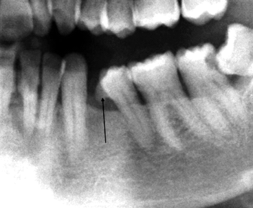
Tartar deposits can be found both in patients with minimal and initial periodontal alterations and in the presence of serious anatomical-radiological situations (Figs. 6.1 and 6.2).
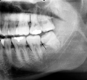
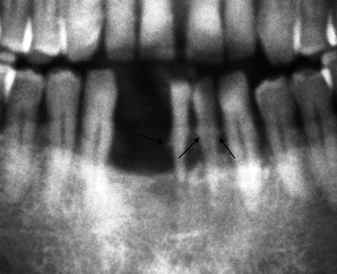

Fig. 6.1
Very thin tartar spicular appositions (indicated by arrows) in the necks with minimal resorption phenomena of the interdental crests

Fig. 6.2
Tartar spicular appositions (indicated by arrows) associated with serious resorption phenomena of the alveolar bone
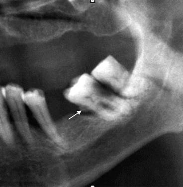
Spicula usually originates at the level of the cementoenamel junction but can, rarely, be localised more distally, in the adjacent radicular surface (Fig. 6.3).
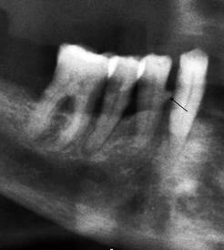

Fig. 6.3
Rough spicula (indicated by arrow) originating at radicular level
Resorption of the Alveolar Bone (Horizontal or Vertical/Angular)
In the natural course of periodontal disease, the first alterations, which occur at the level of the alveolar bone component, are represented by the phenomena of demineralisation and resorption of cortical and cancellous bone. These are an effect of the chemical inflammation agents.
It is known that quantitative evaluations (thickness measurements) and qualitative evaluations (attribution of the skeleton structure to a certain category) are tasks pertaining to CT.
Therefore, OPT turns out to be mainly limited to the morphological judgement of these alterations.
The aforementioned alterations can vary in grade from minimal to serious, the latter of which is characterised by depletion of the alveolar bone.
From a general point of view, the loss of the bone substance can either follow a horizontal or vertical/angular course.
Stay updated, free dental videos. Join our Telegram channel

VIDEdental - Online dental courses


