and Silvio Mazziotti1
(1)
Department of Radiological Sciences, University of Messina, Messina, Italy
Abstract
Inflammatory periapical lesions can be confusing in some aspects due to the lack of pathognomonic radiological images, able to characterise the different pathological entities, and the lack of clinical verification which often characterises the orthopantomography radiological reports. The possibility of having clinical functional information at disposal can be the key factor in precisely marking out the radiological images, which often are nonspecific. It is evident that, in the frequent case of the evaluation of radiological images in a context lacking of clinical information, the radiologist has to describe the picture accurately, without employing terms which subtend complex pathological realities, but describing only elementary alterations (interruption of the lamina dura, periapical lytic lesion, perialveolar osteosclerosis, etc.).
Inflammatory periapical lesions turn out to be complex and can be confusing in some aspects, also for the experts. This is mainly due to the lack of pathognomonic radiological images, able to characterise the different pathological entities, and the lack of clinical verification which often characterises the OPT radiological reports.
Indeed, there is no doubt that the possibility of having clinical functional information at disposal (sensitivity tests, anamnestic confirmation, etc.) can be the key factor in precisely marking out the radiological images, which often are nonspecific.
On the basis of what is mentioned above, it is evident that, in the (very frequent) case of the evaluation of the radiological images in a context lacking of clinical information, the radiologist has to describe the picture accurately, without employing terms which subtend complex pathological realities (e.g. apical granuloma, acute periapical abscess), but describing only elementary alterations (interruption of the lamina dura, periapical lytic lesion, perialveolar osteosclerosis, etc.).
Nosography of Inflammatory Periapical Lesions
As explained above, even if in the majority of cases, the radiological image is to be considered as nonspecific, a brief nosologic overview of the topic seems to be useful (also in order to use a correct terminology).
Alterations of the periapical tissues can be triggered by inflammatory phenomena of endodontic or periodontal origin (Figs. 5.1a, b and 5.2a, b).
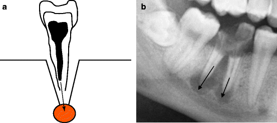
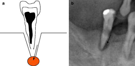

Fig. 5.1
Schematic diagram (a) and radiological representation (b) of periapical lesion of endodontic origin (arrows)

Fig. 5.2
Schematic diagram (a) and radiological representation (b) of periapical lesion of periodontal origin (arrows)
In the first case, the infectious process reaches the periapical region following alterations in the dental pulp, which in turn are caused by penetrating caries or by conditions which cause the contamination of the endodontic content (incongruous treatments, traumatic lesions, etc.)
Conversely, in the second eventuality, the periapical lesion derives from an infection of the periodontal space which represents a predetermined pathway of diffusion (this topic will be treated more extensively in the following chapter).
From the clinical point of view, periapical alterations can be classified as acute, subacute and chronic.
Acute alterations are a consequence of oedematous or necrotic pulpitis, which in most cases, will not give rise to any radiologically recognisable modification.
Conversely, if the pathological process goes further forward, acute periapical abscess originates.
This last, if correctly and promptly treated, can evolve towards healing. Otherwise it can be characterised by a torpid and prolonged course (subacute periapical abscess and chronic periapical abscess). In this case it is followed by possible complications (fistulisation, osteomyelitis, apicolysis, etc.).
Further evolution of the lesion will gradually lead to its progression, with extended destructive phenomena of the alveolar cancellous bone (chronic alveolitis). These may or may not be associated to reactive sclerosis of the surrounding bone trabeculae (osteitis condensans).
Chronic lesions also include the apical granuloma.
This is a lytic lesion of granulomatous nature. Most of the time it is bacterially sterile and it has a scarce clinical appearance (it is often an asymptomatic lesion). However, it is also the expression of an infectious process of endodontic origin.
The presence of epithelial rests (rests of Malassez) in the context of granuloma can influence the cystic evolution, with the consequent formation of a radicular cyst (an entity which will be treated in Chap. 7).
Periapical Abscess
As previously explained, radiological images turn out to be generally nonspecific.
Conversely, the elementary alterations supporting them present a precise pathological significance.
It follows that the medical report will describe those alterations, avoiding the labelling of the lesions with terms which often turn to be imprecise, and even erroneous.
For this reason, the frequent impossibility of discerning a small chronic periapical abscess from an apical granuloma seems to be paradigmatic. This occurs if the morphological element is considered exclusively without any reference to clinical context.
Acute periapical abscess, if not treated by therapy, can appear as a minimal widening of the periapical periodontal space, which, in its early phases, can still be associated to the integrity of the lamina dura (Fig. 5.3a, b).
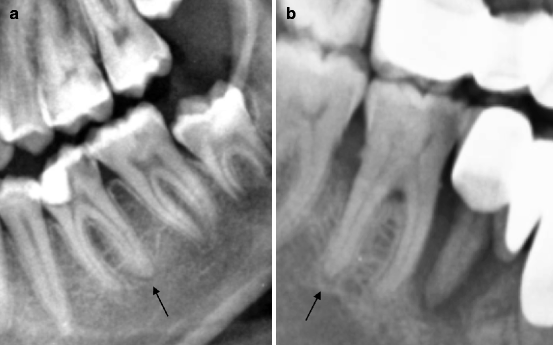

Fig. 5.3
Two cases of minimal and initial periapical inflammatory alteration (indicated by arrows). The periodontal space seems to be widened with integrity of the lamina dura. (a) Secondary caries of 3.7. (b) Caries of 4.6
It seems to be evident that in such cases, the abscess diagnosis is not justified only on the basis of radiological alteration, as analogue modifications of the periodontal space can be supported by inflammatory phenomena. These are constituted only by oedema and cellular infiltrates which are not in purulent evolution.
Further progression of the lytic lesion of the periapical cancellous bone, along with the disappearance of the lamina dura, will create a clear abscessual collection.
In such a phase, the lesion will be characterised by blurred outlines because of the indistinguishable interface between the abscessual collection and the surrounding trabecular bone (Fig. 5.4a, b).
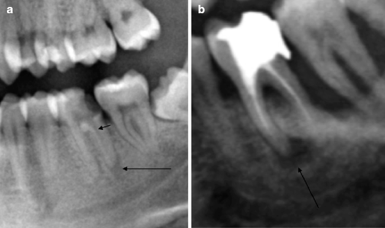

Fig. 5.4
Further progression of the periapical lesions (indicated by arrows) in abscessual direction, with interruption of the lamina dura. (a) Shows penetrating caries of 3.6 with widening of the canal due to endodontic damage (it is indicated by the short arrow). In picture (b) the lesion seems to be more extended and with blurred outlines
From one hand, the subsequent modifications of periapical abscess depend on the efficiency and the timeliness of therapeutic approach (both medical and odontoiatric); from the other hand, they depend on the patient’s immune system capacity.
Indeed, the lesion, in those cases with a slightly aggressive course, will present a thin sclerotic delimitation representing a restraining reaction produced by the adjacent cancellous bone.
In such cases, as will be discussed later, the lesion will turn out to be indistinguishable from the apical granuloma (Fig. 5.5).
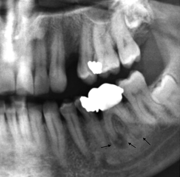

Fig. 5.5
Two periapical lesions (abscesses) in different evolutionary stages, but with phenomena of perifocal sclerosis (indicated by arrows). The lesions are indistinguishable from apical granuloma
Abscesses with major aggresiveness and/or prolonged progression could sometimes be associated with more extended phenomena of osteosclerosis.
This last consideration is extremely indicative of the abscessual nature of the periapical lesion, which obviously has a chronic course (Fig. 5.6).
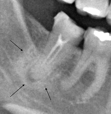

Fig. 5.6
Periapical lesion delimited by a thick vallum of sclerosis of the surrounding spongiosa (indicated by arrows). This element confirms the abscessual nature of the periapical lesion
In those cases in which the resistance capacities of the host bone are not able to contain the further evolution of the morbid process, progressive growth of the abscessual collection occurs. The latter can sometimes reach conspicuous dimensions (Fig. 5.7a–c).
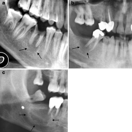

Fig. 5.7
(a–c) Three periapical abscesses (indicated by arrows). In picture (c) the lesion is delimited by a thin band of osteosclerotic reaction
Fistulisation phenomena and massive destruction of the alveolar bone (chronic alveolar abscess) are included within the abscess’ unfavourable evolution.
Stay updated, free dental videos. Join our Telegram channel

VIDEdental - Online dental courses


