and Silvio Mazziotti1
(1)
Department of Radiological Sciences, University of Messina, Messina, Italy
Abstract
In orthopantomography, the evaluation of the masticatory organs’ normal anatomy is different for subjects of paediatric age and adults. Indeed, the first ones present a radiographic case which can be defined as ‘mixed dentition’, in which deciduous dental elements, permanent dental elements and permanent elements in dehiscence coexist all together. Therefore, in paediatric age, considerations of anatomic order and orthopantomography possibilities are completely different from the ones applicable in the study of an adult subject. In order to be more clear on the exposition, we will firstly take into analysis the adult anatomy, that is, the subject with a completed dental development. In the adult, the study of normal anatomy firstly envisages the definition of some general concepts, then subsequently a focused analysis on the following structures: general anatomy of the tooth, anatomy of the support apparatus, anatomy of the mandible and the mandibular canal and anatomy of the surrounding structures (paranasal sinuses, hard palate, nasal cavities, etc.)
In OPT, the evaluation of the masticatory normal anatomy is different for subjects of paediatric age and adults.
Indeed, subjects of paediatric age present a radiographic appearance which can be defined as ‘mixed dentition’, in which deciduous dental elements and permanent dental elements coexist all together.
Therefore, in paediatric age, considerations of anatomic order and OPT possibilities are completely different from the ones applicable in the study of an adult subject.
We will firstly take into analysis the adult anatomy, with a completed dental development.
In the adult, the study of normal anatomy firstly is based on the definition of some general concepts, then subsequently a focused analysis on the following structures:
-
General anatomy of the tooth
-
Anatomy of the support apparatus
-
Anatomy of the mandible and the mandibular canal
-
Anatomy of the surrounding structures (paranasal sinuses, hard palate, nasal cavities, etc.)
Concepts of General Anatomy
The radiographic anatomy of the tooth in the adult, achievable through OPT, does not present the same detail refinement of the radiographic anatomy evaluated through endoral radiograms. This is due to the technical peculiarity of the methodology which favours a global vision rather than a detailed evaluation.
However, even if facing this limitation, dental images achieved through OPT are sufficient to fully express the macroscopical characteristics of the dental element, in the majority of the diagnostic needs.
On the anatomical point of view, it is obviously necessary to take each of the dental typologies (incisors, canines, premolars and molars) into account separately.
In order to easily identify the different dental elements, a classical scheme is considered. It visualises the subdivision of the radiogram in four quadrants which are indicated with progressive numbers (1, 2, 3, 4), from right to left, from bottom up and clockwise.
Each number is associated to the relative dental element of each hemiarch. Number 1 indicates the median incisor, number 2 indicates the lateral incisor, number 3 indicates the canine, etc.
According to this method, for example, the third inferior right molar is indicated with number 4.8, the superior left canine is indicated with number 2.3, etc. (Fig. 2.1).
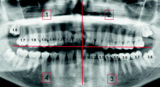

Fig. 2.1
Scheme showing the topographic detection of the dental elements
Such methodology, in addition to facilitating the detection of a specific dental element (in the OPT report), is also extremely useful for evaluating cases with alterations regarding the dental number, both in excess and, more rarely, in defect.
Descriptive and Radiographic Anatomy of the Dental Element
From the structural point of view, the tooth is an architectonical construction which comprises macroscopical and morphofunctional components. They have different purposes; therefore, they are constituted by different tissue types.
Indeed, the tooth is characterised by a visible component called crown and an intraosseous component called root (Fig. 2.2).
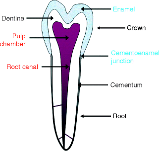

Fig. 2.2
Schematic diagram which illustrates the different morphological and structural components of the tooth
The dental crown represents the different functions of the tooth (shredding, stripping, section) through different morphologies (Fig. 2.3a, b).
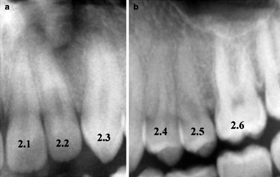

Fig. 2.3
(a) Crown morphology of the median and lateral incisors and of the left canine (2.1, 2.2 e 2.3) aiming at stripping. (b) Crown morphology of the premolars 2.4 and 2.5 and of the first left molar (2.6) aiming at shredding
The root is involved in the support apparatus and in the anchorage of the tooth to the jawbone.
The root end of the tooth is called tooth apex and it is characterised by a very thin hole which gives rise to the vascular-nervous axis.
The dental element, on its whole complex, is constituted by dentin which is an organic substance of mesodermic origin. It is covered by an external sheath called enamel (of ectodermal origin) in the crown and by the cementum (of mesodermic origin) in the root.
Therefore, the dentin is present both in the crown and in the root; it is composed of an organic component produced by the odontoblasts (about 30 %) and by a mineral component (about 70 %) which is composed of hydroxyapatite crystals.
Odontoblasts produce dentine in a dynamic manner both during the tooth’s physiological development (primary dentine) and in pathological conditions (secondary or reactive dentine).
The enamel (produced by a cells called ameloblasts) covers the crown. Therefore, it turns out to be the seat of further mechanical stress; for this reason, it is composed of hydroxyapatite crystals for the 96 %.
Finally, the cementum is also composed of an organic matrix and a mineral component which determines the formation of the so-called mature cementum.
It covers the dentine at the level of the root axis line and is generated by cementoblasts. They are cellular elements placed in the tooth surface which are able to produce cementum for the whole life of the tooth.
Dental elements can be distinguished in monoradicular, biradicular and triradicular teeth.
As a general rule, even if subjected to numerous exceptions, it is possible to establish that incisors and canines of both arches turn out to be always monoradicular teeth. Moreover, all the premolars of the lower arch and the second premolars of the upper arch are monoradicular teeth, too.
Conversely, the first premolars of the upper arch generally have two roots (a vestibular one and a palatal one), while the first and the second molar of the lower arch (which are biradicular teeth, too) are characterised by a mesial and a distal root.
The first and the second molars of the upper arch always turn out to be triradicular teeth (having a mesial, a distal and a palatal root).
Finally, the third molars present a certain variability. They can be both monoradicular and, more rarely, biradicular teeth.
A transition area called neck of tooth or cementoenamel junction exists between the crown and the root. This is the area where the ameloblastic layer, represented by enamel, meets the root surface, which is constituted by the cement.
Inside the tooth, which presents a high radiopacity due to its structure, it is possible to recognise a radiolucency area which exist both in the crown and the root. Such area is, respectively, the expression of the pulp chamber and the root canal (Fig. 2.4).
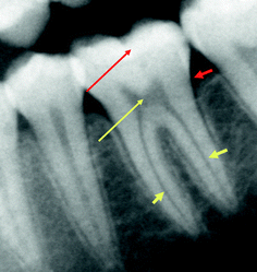

Fig. 2.4
The long red arrow indicates the enamel characterised by maximum opacity. The short red arrow indicates the cementoenamel junction. The long yellow arrow indicates the pulp chamber. The short yellow arrows indicate the radicular canals
The root canal is characterised by very thin horizontal and oblique collateral canals, called lateral canals (however, they are never radiographically visible).
Vascular-nervous elements exist both in the pulp chamber and in the root canal. Together with a connective support (of mesodermic origin) they constitute the dental pulp.
The dental pulp has three purposes: dentine formation (dentinogenesis) and both nutritive and sensorial functions.
To conclude this general analysis of the dental anatomy, it must be remembered that from a topographic point of view, each tooth is characterised by a face which is oriented towards the cheek, called vestibular surface, and another face which is oriented towards the tongue, called lingual surface.
Another face fulfils the mastication process and is particularly augmented in the molars. It is called the occlusal surface as it is influenced, in its morphology, by juxtaposition or occlusal rapports with the dental elements of the opposed arch.
Furthermore, two other faces exist. One face, directed towards the median line, is called mesial surface and another face, opposed to it, is called distal surface. They are interfaces between two dental elements, thereby called interproximal surfaces (Fig. 2.5).
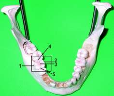

Fig. 2.5
1 Vestibular surface. 2 Lingual (or palatal) surface. 3 Occlusal surface. 4 Distal surface. 5 Mesial surface
As aforementioned, this distinction is also valid for the denomination of the root axis surfaces. Indeed, in a triradicular element, it is possible to distinguish the mesial root from the distal one, in addition to the third root, called palatal. The latter seems to be typically present at the level of the first and the second upper molars and it is not always demonstrable through OPT, for projective reasons.
Indeed, the palatal root develops itself in a different plane with respect to the other roots.
As it is placed farther from the detection plane, it will turn out to be radiographically less defined and rather enlarged (Fig. 2.6a, b).
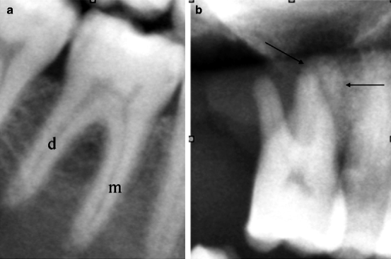

Fig. 2.6
(a) The image shows the biradicular tooth; letter ‘d’ indicates the distal root; letter ‘m’ indicates the mesial root. (b) The image shows the triradicular tooth. The palatal root (indicated by arrows) seems to be enlarged with blurred outlines
It is obvious that the aforementioned expressions of topographic and semantic order are absolutely necessary in order to localise the morbid processes. Therefore, they are propaedeutical to a correct draft of the radiological report.
Descriptive and Radiological Anatomy of the Support Apparatus or Periodontium
The anatomical and functional structure is responsible for the stability of the dental elements, and in final analysis, of the whole masticatory apparatus. It is constituted by the following components: the gingiva, the periodontal ligament (PDL) and the alveolar bone, which in turn is constituted by the spongiosa and the cortical plate (lamina dura). According to some experts, the cementum is part of the alveolar bone, too (Fig. 2.7).
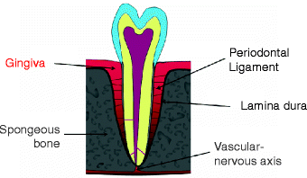

Fig. 2.7
Schematic diagram which represents the periodontium
OPT allows a panoramic evaluation of the normal anatomy and, consequently, of the pathological alterations related only to some of the aforementioned components.
Indeed, it is well known that the evaluation of the gingival apparatus is exclusively of clinical pertinence and also a direct radiographic visualisation of the periodontal ligament is not possible. As a consequence, the state of the periodontal ligament can be indirectly evaluated according to the extent of the periodontal space which, in a normal subject, turns out to be radiographically invisible or scarcely detectable.
Therefore, the radiographic representation of the periodontal apparatus is referred only to the analysis of the alveolar bone, which is constituted by interdental and intraradicular cuspids, with their relative components (spongiosa and lamina dura). Moreover, it is also referred to the evaluation of the periodontal space extent and to the state of the dental roots (Fig. 2.8).
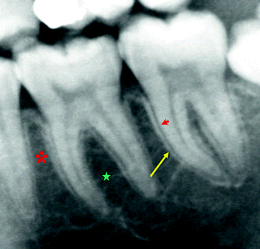

Fig. 2.8
Radiographic representation of the periodontal apparatus. The arrowhead indicates the periodontal space; the yellow arrow indicates the lamina dura; the green star indicates the spongiosa of the interadicular cuspid; the asterisk indicates the spongiosa of the interdental cuspid
The periodontal space is a thin radiolucent line which hosts the namesake ligament, constituted by collagen fibres produced by fibroblasts. Fibroblasts originate from the embryonic cellular elements which are contained in the dental sac.
The elements of the aforementioned ligament-fibrous apparatus (Sharpey’s fibres) are hooked up to the cementum and to the cortical plate of the bundle bone at the level of the alveolar face.
Stay updated, free dental videos. Join our Telegram channel

VIDEdental - Online dental courses


