The kidneys (renal system)
Publisher Summary
This chapter discusses the functional anatomy and histology of the kidneys. The two kidneys lie on either side of the body against the dorsal body wall in the upper part of the abdomen. Each has a ureter attached to the inner curve and these pass down to the upper corners of the triangular-shaped bladder. The bladder is connected to the external body surface by the urethra. All arteries, veins, nerves, and lymphatics enter at the slit-like hilus and pass into the renal sinus, a C-shaped potential space surrounded by the kidney tissue. The functional units of the kidneys are the nephrons. Each kidney contains over a million of these units. The filtered fluid contains all the salts of plasma, glucose, amino acids, and a small amount of protein. Hydrogen carbonate ions are effectively reabsorbed in the proximal tubule. The kidney operates, not by selectively extracting excess and waste products from the blood, but by taking a filtrate of blood and reclaiming from it those materials the body needs to retain. The work involved in the process can be appreciated by considering the amounts of water and solutes reaching the kidney in the circulation as compared with the amounts finally lost in the urine.
Functional anatomy and histology of the kidneys
The two kidneys lie on either side of the body against the dorsal body wall in the upper part of the abdomen. Each has a ureter attached to the inner curve and these pass down to the upper corners of the triangular-shaped bladder. The bladder is connected to the external body surface by the urethra. All arteries, veins, nerves and lymphatics enter at the slit-like hilus and pass into the renal sinus, a C-shaped potential space surrounded by the kidney tissue (Fig. 10.1). The space is potential because it is filled by the structures mentioned and by the renal pelvis with its branches, the major and minor calices. These are the tubes connecting the renal papillae with the ureter. Projecting into the sinus are 8-10 papillae, each of which has at its apex some 18-24 openings of the papillary ducts formed by the fusion of many collecting ducts. In section the kidney has a reddish brown granular-looking outer layer, or cortex, and a red- to grey-brown inner portion, the medulla. The outermost part of the cortex is even in appearance and made up mainly of the proximal and distal parts of the nephrons with some glomeruli. The inner part which dips down into the less even-looking medulla, is marked with lines, or rays, where straight portions of the kidney tubules and blood vessels have been cut longitudinally. The inner cortex contains all parts of the nephron – glomeruli, proximal and distal convoluted tubules, some loops of Henle, and collecting ducts. The medulla presents a rayed appearance because it consists mainly of parallel structures – blood vessels, ascending and descending limbs of loops of Henle, and collecting ducts. Sometimes an inner zone can be distinguished which contains only thin loops of Henle in addition to the collecting ducts and the blood vessels.
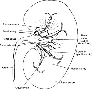
Figure 10.1 A Longitudinal Section Through a Kidney. In the Upper Part a Renal Artery and Its Branches are Shown, in the Lower Part a Renal Vein and Its Branches.
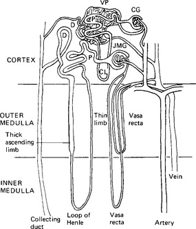
The nephron
The functional units of the kidneys are the nephrons. Each kidney contains over a million of these units. Since the number actually needed is estimated to be of the order of half a million, a generous reserve is available. The nephron is a blind-ended epithelial tube, continuous with the collecting ducts, the papillary ducts, and, through the calices, the ureter (Fig. 10.2). The blind end of the tube, Bowman’s capsule, is wrapped closely around a knot of capillaries, the glomerulus. Each renal corpuscle – the glomerulus together with Bowman’s capsule – is about 100 μm in diameter. From the capsule the nephron continues as the proximal convoluted tubule, a tube lined with a single layer of cuboidal cells with numerous short processes projecting into the lumen. The proximal convoluted tubule coils locally and then extends down a cortical ray to enter the deeper cortex and outer medulla as the relatively thick straight portion which continues as the thin descending limb of the loop of Henle. This second subdivision of the renal tubule is made up of the thin descending limb, the thin segment of the ascending limb, and then the thick segment of the ascending limb which continues on into the distal convoluted tubule. The renal corpuscles in the middle zone of the cortex have loops of varying length which may or may not extend into the medulla and may lack thin segments. The loops entering the medulla from this zone are generally short and thin, in contrast with those originating in corpuscles in the inner zone of the cortex, the juxtamedullary nephrons, which are long and thin. In man these latter constitute only about one eighth of all the nephrons. The thin tubules are made up of only one layer of cells, a thin flattened layer. The ascending limb of the loop of Henle changes abruptly from its thin portion to a thick segment, and this then becomes the beginning of the distal convoluted tubule. From there the distal convoluted tubule passes straight outwards towards its parent corpuscle, and then coils itself in the cortex. There is a transition from cuboidal to columnar cells along its course. The distal convoluted tubule passes close to the afferent arteriole as it enters Bowman’s capsule. Here the arteriolar wall forms a thickened cuff in close approximation to a short specialised zone of the distal convoluted tubule with columnar rather than cuboidal cells. This complex is termed the juxtaglomerular apparatus and is an important organ in the control of kidney function. The distal convoluted tubules finally unite to form the collecting ducts, usually via a short transitional segment. Collecting ducts formed in the outer cortex increase in size as they pass towards and into the medulla, picking up more distal convoluted tubules as they go. They fuse to form the papillary ducts which then open into the calices. The total length of a nephron, excluding the collecting duct, is between 20 and 44 mm. and the average length of a collecting duct is around 22 mm. Since the proximal convoluted tubules are almost two thirds of the length of the nephrons they make up the greater part of the cortex.
Stay updated, free dental videos. Join our Telegram channel
VIDEdental - Online dental courses


