Fig. 3.1
Shows specific ion penetration between glass-ionomer cement and dentine after 2 weeks (Courtesy of Dr. Geoff Knight)
3.1.3 Fluoride Release
EPMA studies have demonstrated in vitro that fluoride released from glass-ionomer cements into demineralized dentine penetrates deeply into the underlying dentine at concentrations of about 5000 ppm (Ngo et al. 2006). Aqueous fluoride concentrations as low as 600 ppm have been shown to inhibit fluoride-resistant Streptococcus mutans bacteria (Brown et al. 1980) (Fig. 3.2).
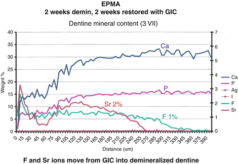
Fig. 3.2
This specimen was demineralized in an demineralizing solution (mimicking caries) onto which an auto-cure glass-ionomer cement restoration was placed. The specimen was examined 2 weeks after placement of the glass-ionomer. EPMA measurement of ion penetration from glass-ionomer cement into demineralized dentine. Note the concentration of fluoride ions at 1 % (about 5000 ppm) to the depth of demineralization, 300 μm into the dentine (Courtesy of Dr. Geoff Knight)
3.1.4 Remineralization
The penetration of strontium, calcium and fluoride from glass-ionomer cements into dentine (Knight et al. 2007a, b) indicates that these ions are available to assist with remineralization of any demineralized dentine remaining beyond the restorative interface.
The combination of dentinal tubular fluid and ion penetration from auto-cure glass-ionomer cements into demineralized dentine creates an environment that predisposes to fluorapatite formation and increase in hardness of the demineralized tooth structure (Fig. 3.3).
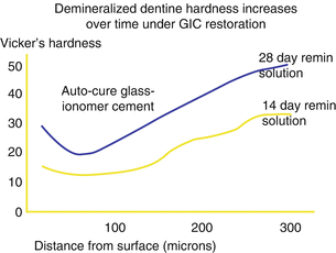
Fig. 3.3
Increase in Vickers hardness of demineralized dentine in a remineralizing solution of artificial saliva (to simulate the oral cavity) under a glass-ionomer cement restoration between 14 and 28 days (Courtesy of Dr. Geoff Knight)
3.1.5 Marginal Caries Protection
Both auto-cure and resin-modified glass-ionomer cements protect the margins of restorations from caries up to depths of 0.25 mm (Knight et al. 2007b; Tantbirojn et al. 2009) while composite resins do not (Fig. 3.4).
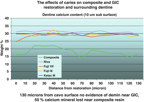
Fig. 3.4
EPMA of subsurface dentine after a 2-week challenge by Streptococcus mutans (simulated chemostat caries study) demonstrating 50 % loss of subsurface calcium adjacent to a composite resin restoration, while there has been no calcium loss from dentine restored with glass-ionomer cements (Courtesy of Dr. Geoff Knight)
3.1.6 Gingival Biocompatibility
Although there is conjecture in the literature about the effects that composite resin and glass-ionomer surfaces have on oral biofilm formation, a survey of the clinical observations of a group of dentists found that gingival inflammation associated with glass-ionomer cement restorations was rarely seen, whereas it was often seen with composite resin restorations (Forsten 1998).
3.1.7 Contouring
Unlike composite resins, amalgams or indirect restorations, glass-ionomers are relatively soft and easy to contour with either low- or high-speed instruments, especially in difficult access areas or at cervical margins.
3.1.8 Aesthetics
The aesthetics of the newer auto-cure glass-ionomer cements are approaching that of resin-modified glass-ionomer cements. In the aesthetic zone, requiring high aesthetics, composite resins are better suited. However, beyond this zone, most clinicians find that the aesthetics of glass-ionomer cements meet their patient’s requirements.
3.2 Limitations of Glass-Ionomer Cements
3.2.1 Wear Resistance
The low wear resistance of glass-ionomer cements is often cited as a reason to exclude them as an occlusal restorative material. While the surface wear of resin-modified glass-ionomer cements clinically is significant, the restorative auto-cure glass-ionomer cements have an excellent record of low occlusal wear and marginal integrity, providing that they are not placed over occlusal surfaces that involve centric stops (Knight 1992; Lazaridou et al. 2015).
3.2.2 Buffering Oral Acids
Glass-ionomer cements act as buffers to changes in oral pH that will cause their slow degradation in areas where saliva is unable to wash oral acids away. This can result in the surfaces of the glass-ionomer cements being degraded and lost (Nicholson et al. 2000).
In such circumstances, when glass-ionomer cement and composite resin are placed to form an “open-sandwich” restoration, the glass-ionomer cement component of the restoration can be “washed out” to create the effect of proximal recurrent caries. This is possibly the reason why clinicians suggest that they observe proximal caries associated with these restorations (Tyas 2005). While caries may not be a problem, food packing certainly is, and the use of “open-sandwich” restorations should be discouraged as a clinical procedure and a “closed-sandwich” restoration should be placed instead (Figs. 3.5 and 3.6).
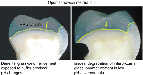
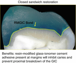
Fig. 3.5
Demonstrates the potential consequences of interproximal plaque acids on the glass-ionomer cement in an “open-sandwich” restoration (Courtesy of Dr. Geoff Knight)
Fig. 3.6
Shows a “closed-sandwich” proximal restoration that prevents loss of proximal glass-ionomer cement (Courtesy of Dr. Geoff Knight)
3.2.3 Residual HEMA (2-Hydroxyethylmethacrylate)
Resin-modified glass-ionomer (RMGIC) restorations have relatively good aesthetics. However, they should be limited to shallow restorations, away from occlusal loads as they have poor wear resistance, e.g. in cervical restorations.
Furthermore, these materials are quite opaque compared to composite resins, and there is limited penetration of light to the base of RMGIC restorations during photo-curing that can leave unpolymerized HEMA remaining at the restorative interface. This predisposes to water uptake from the tooth into the restoration and penetration of unpolymerized HEMA from the restoration into the dentinal tubules and eventually into the pulp (Watson 1997).
Light-cured resin-modified adhesives and luting agents, however, are applied as thin films over tooth surfaces that will result in much higher levels of polymerization of the HEMA upon photo-curing.
3.3 Clinical Applications
3.3.1 Surface Preparation
Glass-ionomer cements adhere as a relatively weak ionic bond (about 2.5 MPa) to any clean tooth surface irrespective if it is enamel, dentine or cementum and sound or carious (Lenzi et al. 2013). The bond strength may be enhanced by removing the surface bioload using either conditioning for 10 s with 20 % polyacrylic acid or etching for 5 s with 37 % phosphoric acid (Van Meerbeek et al. 2003).
3.3.2 Condition or Etch?
Traditionally, manufacturers have instructed clinicians to condition teeth with 20 % polyacrylic acid for 10 s prior to placing glass-ionomer cements and have shown a slightly superior bond achieved in vitro with conditioning compared to etching. In addition, the conditioner contains the same components as the liquid used in glass-ionomer cements, and hence, any residue should not interfere with the bonding process; on the other hand, it is said that etching removes mineral content from dentine that reduces the bond strength (McLean 1992).
Much of this published work has been carried out in vitro, and there are significant clinical differences when applied to the oral environment.
Firstly, most hand-pieces are oiled prior to autoclaving, resulting in a spray of oil over the tooth surface from the hand-piece during cavity preparation. The bond strength of dentine surfaces contaminated with oil and conditioned with polyacrylic acid is half that of a non-contaminated surface (Matos et al. 2008). However, etching removes the oil from the dentine surface; dentine with oil contamination that has been etched has the same bond strength as etched non-contaminated dentine (Matos et al. 2008).
Furthermore, a recent paper has shown that RMGIC bonding agents are equally effective, irrespective of whether they are etched or conditioned (Hamama et al. 2014).
Finally, as the fluoride ions from a glass-ionomer cement penetrate the etched dentine surface and combine with dentinal tubular fluid, the dentine will remineralize to form a caries-resistant layer of fluorapatite.
3.4 Auto-Cure or Resin-Modified Glass-Ionomer Cements?
Auto-cure glass-ionomer cements are better suited as restorative materials than resin-modified glass-ionomers as they have better occlusal wear and do not have the problems associated with residual HEMA found at the base of resin-modified glass-ionomer cement restorations.
3.5 Auto-Cure Glass-Ionomer Cements
3.5.1 Fissure Protection
The use of composite resins to fissure seal occlusal surfaces deemed to be at risk from caries has long been advocated by many in the dental profession.
As teeth form in a “biological soup”, when they first erupt into the mouth, the hydroxyl apatite crystals are contaminated with carbonate groups that make them more prone to acid breakdown at a higher pH than teeth that have been erupted for some time. After eruption, the outer layers of apatite are subjected to a series of demineralization and remineralization cycles, which in the presence of fluoride, will form a layer of fluorapatite crystals that require a lower pH for demineralization and hence are more caries resistant than a newly erupted tooth (Chow and Vogel 2001).
Sealing a fissure with composite resin on a recently erupted tooth prevents the transformation from carbonated apatite to fluorapatite, leaving the tooth potentially more susceptible to caries if the seal was lost. A tooth that has been in the mouth and subjected to multiple demineralization and remineralization cycles will be far more resistant to microbial attack.
Protecting a fissure with auto-cure glass-ionomer cement has the benefit of, firstly, demineralizing the outer apatite crystals due to the low pH of the uncured glass-ionomer. Secondly, after curing when the pH returns to neutral and in the presence of a high concentration of fluoride ions, demineralized apatite crystals will remineralize as fluorapatite.
Eventually, when the glass-ionomer has worn from the surface, the remaining enamel will be able to resist caries attack as well, if not better, than a mature tooth that has been subjected to multiple remineralizing cycles in a high-fluoride environment.
3.5.1.1 Technique
-
Etch the surface to be protected for 5 s with 37 % phosphoric acid, wash and dry thoroughly.
-
Isolate the tooth with cotton rolls.
-
Apply an auto-cure glass-ionomer over the surface and “puddle” into the fissures with a disposable mini-brush.
-
Place a 3 x 2 cm film of a “freezer bag” over the surface and have the patient close in maximum intercuspation. This will force the glass-ionomer into the fissures and maximize the amount that can exist within the occlusion, substantially increasing the area to be protected and the time the glass-ionomer will remain upon the occlusal surface before being worn away (Fig. 3.7).
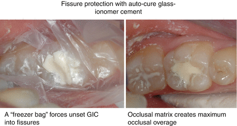 Fig. 3.7Placement of a 3 × 2 cm piece of freezer bag over a glass-ionomer cement when fissure sealing will force the cement into the fissures and defines the occlusal envelope allowing the maximum amount of glass-ionomer cement coverage of the occlusal surface (Courtesy of Dr. Geoff Knight)
3.5.2 Management of Cervical Hypersensitivity
The low-viscosity auto-cure glass-ionomer cements are well suited for protection from cervical hypersensitivity by isolating the tooth surface from the oral environment and releasing fluoride that may well further help encourage desensitization.
3.5.2.1 Technique
-
Clean the surface to be treated ideally with 37 % phosphoric acid for 5 s; depending upon the level of sensitivity, this may require prior administration of a local anaesthetic.
-
Isolate the area with cotton rolls.
-
Mix the low-viscosity auto-cure glass-ionomer and place the unset material on a mixing pad.
-
Using a small disposable mini-brush, paint over the relevant areas identified with the hypersensitivity. Distribution of the glass-ionomer cement may be aided by blowing air gently into the interproximal spaces.
-
Allow the glass-ionomer to cure.
-
A second application of the glass-ionomer may occasionally be required.
3.5.3 Luting Cements
Both auto-cure and resin-modified glass-ionomer cements can be used as luting cements. They are radiopaque and have reasonable adhesion (about 10 MPa) and a relatively high-fluoride release that protects margins from recurrent caries (Tantbirojn et al. 2009).
Auto-cure luting cements have a film thickness of about 20 μm and resin-modified luting cements as low as 10 μm.
Auto-cure luting cements are suitable for metal-based restorations or posts. Resin-modified luting cements have similar properties and benefit from a controlled setting time.
Resin-modified luting cements will bond to both metallic and ceramic surfaces at bond strengths of about 7 MPa. Resin-modified luting cements are best applied to translucent restorations that will facilitate photo-cure polymerization of the HEMA within the cement.
3.6 Auto-Cure Glass-Ionomer Cement as a Restorative Material
The choice of the type of restoration technique depends upon the site of the lesion and the state of the tooth to which it is to be applied. If the lesion occurs in a region of a tooth where the surrounding cusps are well supported and not involving a centric stop, a wear-resistant glass-ionomer cement restoration may be used. When adjacent cusps are undermined and susceptible to occlusal loads, or if the centric stops occur on the occlusal surface, then a composite resin or combined composite resin/glass-ionomer cement restoration may be more appropriate (Figs. 3.8 and 3.9).
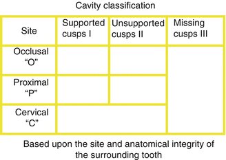
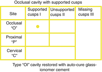
Fig. 3.8
This table, developed by the author, shows a classification for restorations based upon the site of a lesion and the anatomical integrity of the surrounding tooth structure; i.e. if the surrounding cusps are supported and centric stops not involved, then a wear-resistant auto-cure glass-ionomer cement restoration is indicated: if surrounding cusps are not supported, then a composite resin or glass-ionomer composite “sandwich” restoration is required (Courtesy of Dr. Geoff Knight)
Fig. 3.9
Traditional Class I cavity: this classification is for a small occlusal cavity with well-supported surrounding cusps suitable for placing a wear-resistant auto-cure glass-ionomer cement. OI-type cavity (Courtesy of Dr. Geoff Knight)
3.6.1 Occlusal Restorations with Supported Cusps: OI-Type Cavity
These are small cavities that do not involve centric stops and can be placed using auto-cure glass-ionomers.
3.6.1.1 Technique
-
Cavity preparation is completed by preparing a moat around the dentino-enamel junction using a size no. 3 round slow-speed bur to aid retention and assure a biological seal into sound dentine. This acts as a further preventive measure against recurrent caries.
-
After cavity preparation, etch the preparation for 5 s with 37 % phosphoric acid (to remove hand-piece oil and other debris), wash and dry with oil-free air.
-
Isolate with cotton rolls.
-
Place the glass-ionomer cement into the cavity (preferably with a capsule) from the base of the cavity upwards to minimize air inclusions and to slightly overfill the cavity.
-
Place a 3 x 2 cm piece of “freezer bag” over the restoration and ask the patient to close into maximum intercuspation until the glass-ionomer cement cures. This forces the glass-ionomer cement into the cavity and creates an occlusal matrix that minimizes contouring of the cured restoration.
-
In small cavities, the only adjustment required can be done with a sharp excavator.
-
In larger restorations that involve more of the occlusal surface, it may be necessary to contour the inclined planes of the restoration parallel to those of adjacent teeth to compensate for lateral and protrusive movements of the mandible during mastication
-
Covering the restoration with an isolating varnish film is a matter of the clinician’s choice (Fig. 3.10).
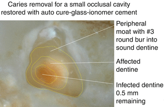 Fig. 3.10Cavity preparation. Remove caries to just above the infected dentine layer as shown, leaving a 0.5 mm layer of carious dentine at the base of the preparation surrounded by affected (remineralizable) dentine. Prepare a moat around the periphery of the cavity into sound dentine using a no. 3 round bur (Courtesy of Dr. Geoff Knight)
3.6.2 Occlusal Restorations with Unsupported Cusps: OII-Type Cavity
These cavities involve more of the occlusal surface such that cusps are either unsupported or involve centric stops. Glass-ionomer cement restorations do not have the shear strength to support these cusps and are subject to high levels of wear on surfaces involving centric stops. In such circumstances, the preferred restorative technique is a “sandwich” technique, described by John McLean, where glass-ionomer cement replaces dentine and composite resin replaces enamel (McLean 1992) (Fig. 3.11).
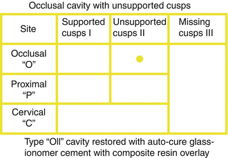
Fig. 3.11
Traditional Class I cavity: this classification is for a larger occlusal cavity when the surrounding cusps are unsupported or the restoration involves a centric stop indicating a composite resin or glass-ionomer composite “sandwich” restoration. OII-type cavity (Courtesy of Dr. Geoff Knight)
3.6.2.1 Technique
-
Caries removal is similar as for a small occlusal restoration: leave 0.5 mm layer of carious dentine and prepare a moat into sound dentine around the dentino-enamel junction using a no. 3 round bur.
-
As the cusps are unsupported, it is advisable to protect them using an occlusal overlay preparation with a high-speed large round diamond bur.
-
After cavity preparation, etch the preparation for 5 s with 37 % phosphoric acid (to remove hand-piece oil and other debris), wash and dry with oil-free air.
-
Isolate with cotton rolls.
-
Place the glass-ionomer cement into the cavity (preferably using a capsule), filling from the base of the cavity upwards to minimize air inclusions up to the dentino-enamel junction.
-
Prepare a resin-modified glass-ionomer adhesive bond and either wait until the auto-cure glass-ionomer has set or place the bond directly over the uncured glass-ionomer and the exposed cavosurface margin walls (Knight et al. 2006). (This procedure is called “co-curing” and discussed later within this chapter.)
-
Insert an increment of composite resin over the freshly set or unset glass-ionomer to slightly overfill the cavity.
-
Burnish the margins with a ball burnisher.
Stay updated, free dental videos. Join our Telegram channel
VIDEdental - Online dental courses


